The process of aging plays a significant role in the steady reduction of how well the brain works and is connected to diseases such as dementia, continuous cognitive decline, and issues with memory. Research suggests that fucoidan has the potential to ameliorate cognitive impairment.
This blog post aims to introduce and discuss a research study carried out by Yan Xu et al. titled “Fucoidan Improves D-Galactose-Induced Cognitive Dysfunction by Promoting Mitochondrial Biogenesis and Maintaining Gut Microbiome Homeostasis” which aimed to elucidate the protective effects of fucoidan against D-galactose (D-gal)-induced cognitive dysfunction and the underlying mechanisms.
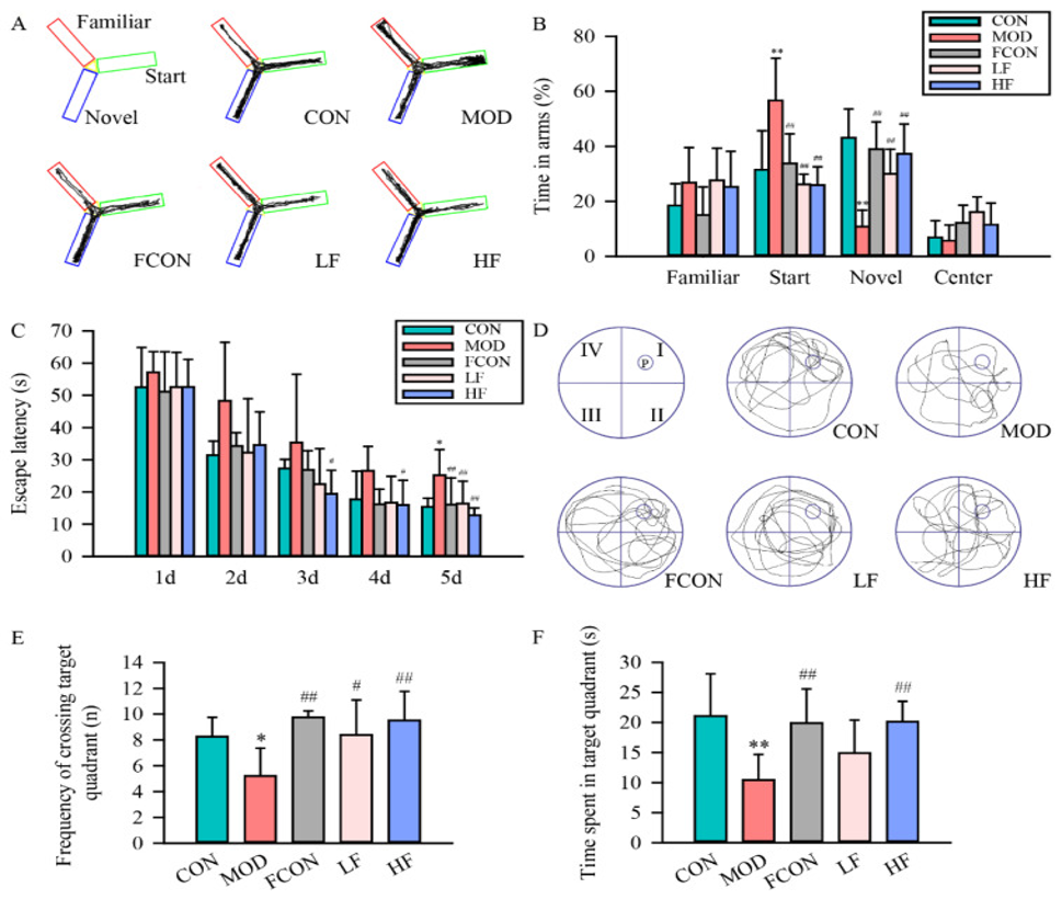
A Y-maze was used to assess spatial recognition memory in animals, and the protective effect of fucoidan against spatial recognition memory impairment induced by D-galactose (D-gal) administration was investigated by observing the rats’ exploration of the “start arm” and “novel arm.” Figure 1A shows a schematic of the Y-maze and typical movement trajectories of the rats within it, while Figure 1B displays the time spent in each arm. In comparison with the Control group (CON), the Model group (MOD) rats allocated significantly more time to the start arm and significantly less time to exploring the novel arm. In contrast to the MOD group, the fucoidan-treated group (FCON) showed reduced time spent in the start arm and increased time exploring the novel arm; no significant difference was observed between the FCON and CON groups.
The Morris water maze (MWM) test was used to evaluate spatial learning and memory function. As shown in Figure 1C, the results indicated that escape latency (the time required to reach the platform) gradually decreased over the training days across all groups. Compared to the CON group, the MOD group exhibited significantly longer escape latency during the final hidden-platform trial. On the other hand, the fucoidan-treated group demonstrated a significantly reduced escape latency in comparison to the MOD group.
Figures 1D–F illustrate typical movement trajectories, the number of crossings into the target quadrant, and the time spent in the target quadrant during the MWM probe test. The results revealed clear cognitive impairment in the MOD group; compared to the CON group, the MOD group showed significant reductions in both the number of crossings into the target quadrant (Figure 1E) and the time spent in the target quadrant (Figure 1F). The fucoidan-treated group exhibited significantly higher values for both the number of crossings into the target quadrant (Figure 1E) and the time spent in the target quadrant (Figure 1F) compared to the MOD group. There was no notable difference observed between the FCON and CON groups.
Concerning the histopathological alterations in the hippocampal CA1 and DG areas identified by HE staining, CA1 pyramidal cells and DG granule cells in the CON and FCON groups showed clear nuclear regions and visible nuclei, with no notable pathological changes detected. In contrast, cells in the hippocampal CA1 and DG regions of the MOD group showed disordered arrangement, nuclear pyknosis, and nuclear hyperchromasia. Compared to the MOD group, the fucoidan-treated group exhibited attenuated pathological changes in the cells of the CA1 and DG regions.
Nissl staining results revealed that, compared to the CON group, cells in the hippocampal CA1 and DG regions of the MOD group exhibited evident damage, indistinct nuclear membranes, blurred nucleoli, and nuclear hyperchromasia. Semi-quantitative scoring used to compare histopathological changes across the groups indicated that the number of normal neurons was significantly lower in the MOD group than in the CON group. The number of normal neurons increased and neuronal damage was lessened by fucoidan.
Oxidative stress and inflammation play crucial roles in aging. To investigate whether fucoidan could alleviate D-galactose (D-gal)-induced oxidative stress damage, we first measured reactive oxygen species (ROS) levels in each group using the DCFH-DA probe; the results showed that probe fluorescence intensity was significantly higher in the MOD group compared to the CON group, whereas it was significantly lower in the HF group compared to the MOD group.
Furthermore, using detection kits, we measured the activities of redox-related enzymes and the levels of malondialdehyde (MDA)—an end-product of lipid peroxidation—in rat brain tissue; the results indicated that CAT and SOD activities were significantly lower in the MOD group compared to the CON group. In contrast, CAT and SOD activities in the HF group were significantly higher than those in the MOD group.
Compared to the CON group, GSH-Px activity was significantly reduced in the MOD group but significantly increased in both the LF and HF groups. While MDA levels were significantly elevated in the MOD group compared to the CON group, fucoidan treatment reduced MDA levels; notably, the HF group exhibited significantly lower MDA levels than the LF group.
No significant differences were observed between the FCON group and the CON group regarding probe fluorescence intensity or levels of CAT, GSH-Px, SOD, and MDA in rat brain tissue. To examine whether fucoidan could suppress D-galactose-induced inflammation, we measured levels of inflammatory markers (LPS, TNF-α, IL-6, and IL-1β) in each group; the results showed that levels of these markers were significantly elevated in the MOD group compared to the CON group, whereas significant reductions were observed in the fucoidan-treated groups compared to the MOD group. Additionally, levels of LPS, TNF-α, and IL-1β were significantly lower in the HF group compared to the LF group.
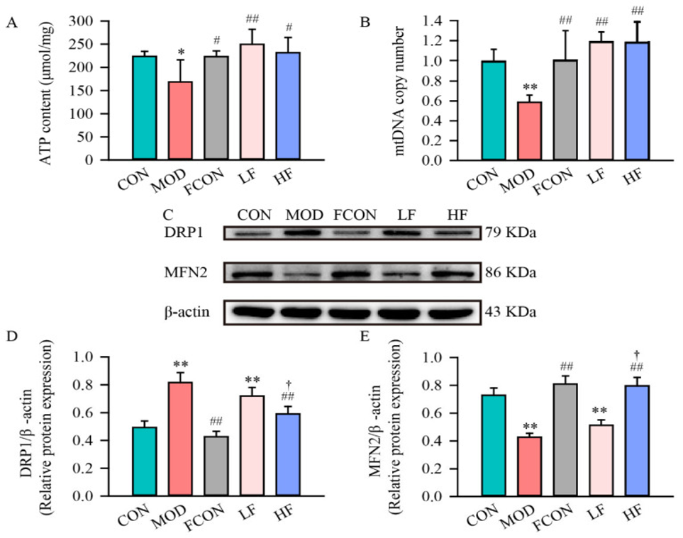
Under conditions of oxidative stress, mitochondria become targets of reactive oxygen species (ROS), leading to mitochondrial dysfunction. To investigate the effects of D-gal (D-galactose) on mitochondrial function in the rat hippocampus, we evaluated ATP content (Fig. 2A) and mtDNA copy number (Fig. 2B). The results showed that hippocampal ATP content was significantly lower in the MOD group compared to the CON group (Fig. 2A); however, in the LF and HF groups, hippocampal ATP content recovered to levels comparable to those of the control group (CON group) (Fig. 2A).
Furthermore, the mtDNA copy number was significantly reduced in the MOD group compared to the CON group (Fig. 2B). Following fucoidan administration, the mtDNA copy number recovered to a level equivalent to that of the CON group (Fig. 2B).
In neurons, an imbalance between fission and fusion can lead to impairments in mitochondrial structure and function; therefore, we measured the expression levels of DRP1 and MFN2 in hippocampal tissue using Western blotting (Fig. 2C). The data indicated that the expression level of the fission-related protein DRP1 was significantly elevated in the MOD group (Fig. 2D), while the expression level of the fusion-related protein MFN2 was significantly reduced in the MOD group compared to the CON group (Fig. 2E). Additionally, in the HF group, DRP1 expression levels were significantly lower (Fig. 2D) and MFN2 expression levels were significantly higher (Fig. 2E) compared to the MOD group.
In addition, we examined the effect of fucoidan on DRP1 protein expression in hippocampal tissue using immunofluorescence staining. The results suggested that DRP1 was primarily localized in the cytoplasm of cells within the hippocampal CA1 and DG regions. DRP1 fluorescence intensity significantly rose in the hippocampal CA1 and DG regions of the MOD group compared to the CON group, but administering a high dose of fucoidan decreased these intensities in the respective regions.
To investigate the relationship between mitochondrial dysfunction and mitochondrial biogenesis, we examined the expression of PGC-1α, NRF1, and TFAM in the hippocampal tissue of each group. The results showed that the protein levels of PGC-1α, NRF1, and TFAM were significantly reduced in the MOD group compared to the CON group. Fucoidan treatment restored the reduced expression of PGC-1α, NRF1, and TFAM in the hippocampal tissue of D-gal-treated rats.
Recent studies suggest that the APN-AMPK-SIRT1 pathway may be involved in regulating mitochondrial biogenesis via the upstream expression of PGC-1α. Therefore, we examined the expression of APN, AMPKα, p-AMPKα (Thr172), and SIRT1 in the hippocampal tissue of each group. The results demonstrated that the expression of APN, the p-AMPKα (Thr172)/AMPKα ratio, and SIRT1 in the hippocampus was significantly lower in the MOD group than in the CON group; furthermore, fucoidan administration increased the expression of SIRT1 and APN, as well as the p-AMPKα (Thr172)/AMPKα ratio.
Additionally, using immunofluorescence to detect the localization and expression intensity of SIRT1 in hippocampal tissue, we found that SIRT1 expression was present in both the nuclei and cytoplasm of cells in the CA1 and DG regions. Moreover, the fluorescence intensity of SIRT1 in the hippocampal CA1 and DG regions was significantly lower in the MOD group compared to the CON group. Fucoidan administration resulted in an increase in SIRT1 fluorescence intensity.
One potential mechanism underlying the anti-inflammatory effects of fucoidan involves the suppression of Firmicutes and the increase of Bacteroidota. Compared to the MOD group, the fucoidan-treated group exhibited a decrease in the abundance of unclassified Lachnospiraceae and an increase in that of unclassified Muribaculaceae. It has been suggested that Muribaculaceae may regulate oxidative stress and inflammation via the gut microbiota-gut-brain axis, potentially modulating cognitive function in patients with Alzheimer’s disease (AD). Furthermore, LDA score analysis revealed that Akkermansia was a predominant bacterial genus in the HF group. Akkermansia has been reported to potentially ameliorate intestinal disorders and extend the healthspan of aging mice; this may be linked to the cognitive-protective effects of fucoidan. The study concludes that fucoidan can decrease inflammation and oxidative stress, relieve mitochondrial dysfunction, and ameliorate cognitive impairment caused by D-galactose (D-gal) by influencing the structure and composition of gut microbiota in rats.
These results demonstrate that fucoidan can relieve cognitive problems induced by D-galactose through the regulation of the APN-AMPK-SIRT1 signaling pathway and the homeostasis of gut microbiota, ultimately reducing oxidative stress and inflammation and improving mitochondrial function. Hence, fucoidan has the potential to be used as a functional food for the prevention of cognitive impairment linked to aging.
Source: Nutrients. 2024 May 17;16(10):1512. doi: 10.3390/nu16101512