The activation of hypoxia-inducible factor-1 (HIF-1), which is prompted under hypoxic conditions, plays a critical role in promoting tumor angiogenesis, proliferation, and metastasis. While low-molecular-weight fucoidan (LMWF) derived from brown algae exhibits anti-cancer properties, it remains unclear whether LMWF inhibits hypoxia-induced angiogenesis in bladder cancer cells or what the underlying molecular mechanisms might be. This blog post introduces the study “Low Molecular Weight Fucoidan Inhibits Tumor Angiogenesis through Downregulation of HIF-1/VEGF Signaling under Hypoxia” by Meng-Chuan Chen et al., which investigated whether LMWF inhibits hypoxia-induced H2O2 production, HIF-1 accumulation and transcriptional activity, vascular endothelial growth factor (VEGF) secretion, and cell migration and invasion in human bladder cancer cells (T24 cells) under hypoxic conditions.
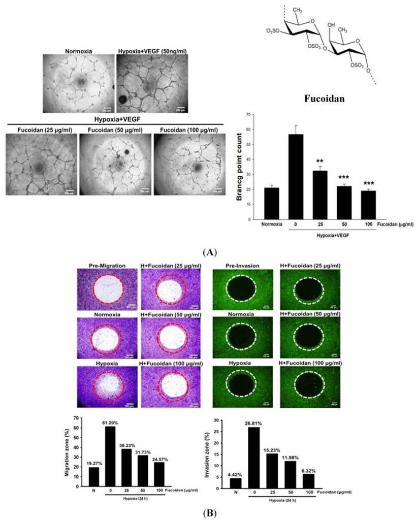
First, they demonstrated whether LMWF inhibits hypoxia-induced angiogenesis in human umbilical vein endothelial cells (HUVECs) and the migration and invasion of human bladder cancer cells (T24 cells). As shown in Figure 1A, LMWF inhibited hypoxia- and VEGF-induced capillary-like structure formation in HUVECs in a dose-dependent manner. In contrast, it did not affect angiogenesis under normoxic conditions. These results suggest that the anti-angiogenic effect of LMWF is hypoxia specific. Since angiogenesis is essential for tumor metastasis, we evaluated the effect of LMWF on the migration and invasion of T24 cells. LMWF treatment significantly inhibited their migration and invasion under hypoxic conditions (Figure 1B).
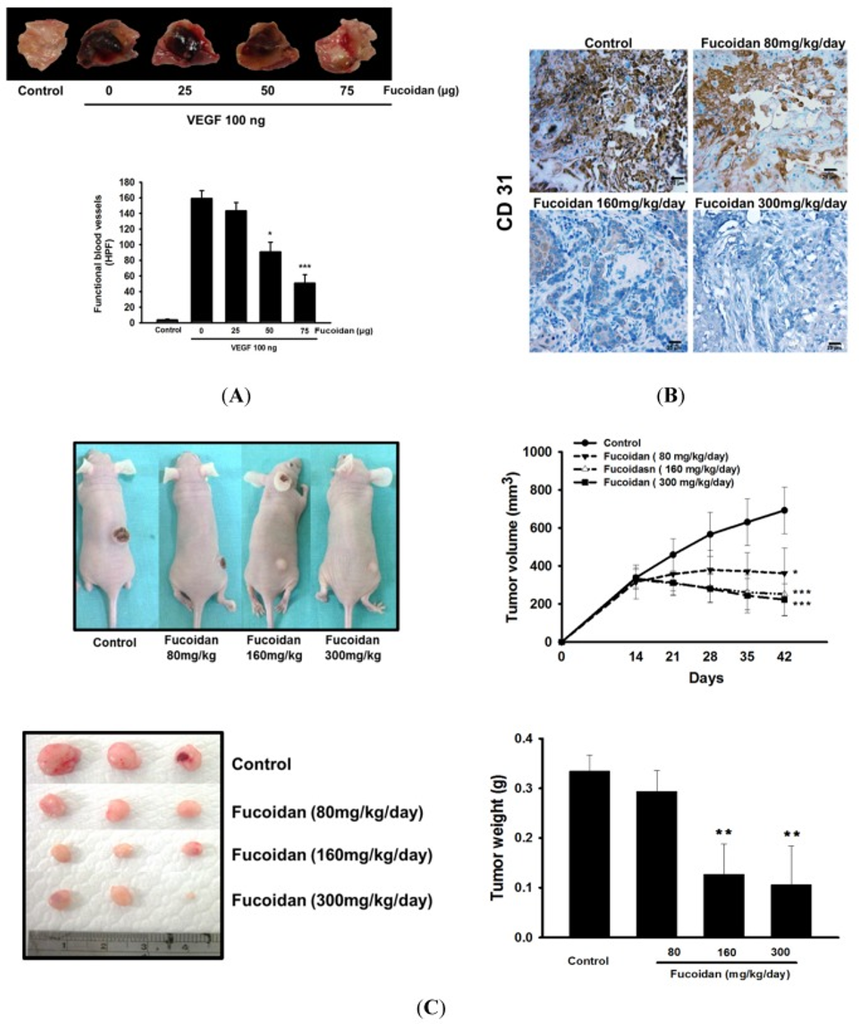
Next, they investigated whether LMWF inhibits angiogenesis and tumor growth *in vivo*. In Matrigel plugs containing VEGF, the control group (untreated tumor-bearing mice) exhibited a markedly dark appearance due to engorgement with numerous intact red blood cells, indicating the formation of functional blood vessels (Fig. 2A). Consistent with this, the level of CD31 (a specific marker for endothelial cells) in the tumor tissue was significantly elevated (Fig. 2B). LMWF administration inhibited *in vivo* angiogenesis in a dose-dependent manner, as evidenced by a reduction in the number of blood vessels within the Matrigel plugs and the number of CD31-stained capillaries within the tumors. Furthermore, tumor size and weight were significantly reduced in LMWF-treated mice compared to the control group (Fig. 2C). Additionally, LMWF treatment did not cause weight loss in the mice, indicating that LMWF is safe at the doses used.
Western blot analysis revealed that exposing T24 cells to hypoxic conditions for 8 hours resulted in a marked increase in HIF-1α nuclear translocation, HIF-1 transcriptional activity, hydrogen peroxide (H2O2) production, and VEGF expression and release, compared to T24 cells under normoxic conditions. However, these hypoxia-induced changes were dramatically suppressed by LMWF. Consequently, LMWF inhibits hypoxia-induced HIF-1α activation, reactive oxygen species (ROS) production, and VEGF release in T24 cells.
In T24 cells, hypoxia-induced stimulation led to increased VEGFR2 phosphorylation and activation, a process that was potently inhibited by LMWF. Previous studies have shown that the PI3K/AKT/mTOR signaling pathway—downstream of activated VEGFR2—can promote HIF-1 protein synthesis by phosphorylating protein translation regulators such as ribosomal p70S6 kinase (p70S6K1) and 4EBP-1; in this study, the hypoxia-induced hyperphosphorylation of AKT, mTOR, p70S6K, and 4EBP-1 in T24 cells was significantly suppressed by LMWF without affecting the total protein levels of these kinases. To further investigate the role of PI3K/AKT/mTOR signaling in the induction of HIF-1α and VEGF, the mTOR inhibitor rapamycin and the PI3K/AKT inhibitor wortmannin were administered. The results revealed that both rapamycin and wortmannin inhibited hypoxia-induced HIF-1α expression and VEGF secretion, suggesting that this pathway plays a crucial role in the transcriptional regulation of HIF-1α and VEGF. These findings indicate that LMWF inhibits the signaling pathway regulating HIF-1 in T24 cells.
In the same way, the increases in nuclear HIF-1 protein levels and VEGF expression observed in tumor tissues were significantly suppressed by LMWF treatment compared to untreated tumor-bearing mice. This suggests that the anti-angiogenic and anti-tumor effects of LMWF are associated with the downregulation of HIF-1 and VEGF expression; specifically, LMWF suppresses HIF-1α and VEGF expression in tumors.
Based on these findings, the study demonstrated that LMWF inhibits hypoxia-induced H2O2 production, HIF-1 accumulation and transcriptional activity, VEGF secretion, and cell migration and invasion in hypoxic human bladder cancer cells (T24 cells); furthermore, the anti-angiogenic effect of LMWF on bladder cancer under hypoxic conditions may be associated with the inhibition of the HIF-1/VEGF-regulated signaling pathway.
Source: Mar Drugs. 2015 Jul 17;13(7):4436–4451. doi: 10.3390/md13074436