
In this blog post, the study “Cytotoxic Effects of Fucoidan Nanoparticles against Osteosarcoma” by Ryuichiro Kimura et al. is introduced. The study examined how the biological activity of fucoidan varies with its size by comparing the cytotoxic effects of unmodified fucoidan and fucoidan-loaded lipid nanoparticles on osteosarcoma cells, using both laboratory (in vitro) and live animal (in vivo) models.
First, they investigated the cytotoxic effects of fucoidan on osteosarcoma cells. To elucidate the structure of the prepared SLP-PC70 liposomes, they performed freeze-fracture electron microscopy (FFEM) observation. The liposomes were confirmed to be small unilamellar vesicles (SUVs) with a particle size of approximately 100 nm.
They also evaluated the cytotoxic effects of nanoparticle fucoidan on the human osteosarcoma cell line 143B using the water-soluble tetrazolium (WST)-8 assay. Nanoparticle fucoidan reduced cell viability in a dose- and time-dependent manner.
Apo2.7 specifically detects the 38-kDa mitochondrial membrane antigen 7A6, which is expressed on the outer mitochondrial membrane during apoptosis; as demonstrated by Apo2.7 staining, nanoparticle fucoidan induced apoptosis in 143B cells in a dose- and time-dependent manner.
Next, they examined the effect of HMWF (high-molecular-weight fucoidan) on apoptosis induction. At a concentration of 2 mg/mL, the apoptosis-inducing activity of native fucoidan was lower than that of nanoparticle fucoidan. These findings indicate that nanoparticle fucoidan has greater activity than native fucoidan in triggering apoptosis in cultured osteosarcoma cells. Caspases are a group of cysteine proteases that play a crucial role in the apoptotic cascade; pre-incubation of 143B cells with the pan-caspase inhibitor Z-VAD-FMK for 60 minutes before the addition of nanoparticle fucoidan significantly inhibited the reduction in cell viability. According to these findings, nanoparticle fucoidan causes apoptosis by activating the caspase pathway.
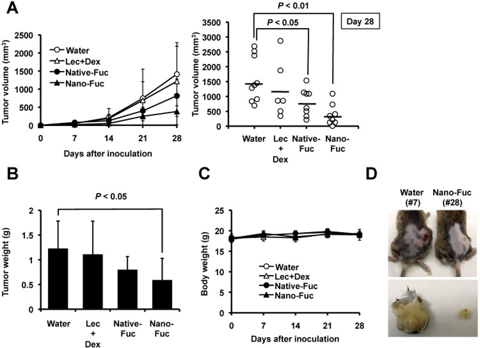
They studied the impact of fucoidan on tumor growth in vivo to explore its potential in treating osteosarcoma. After implanting murine osteosarcoma LM8 tumor cells into the backs of C3H mice, they orally administered either nanoparticle-formulated fucoidan or unprocessed fucoidan at a dose of 100 mg/kg/day. Control mice received only water. Administration of both unprocessed and nanoparticle-formulated fucoidan resulted in a significant reduction in tumor volume in LM8-implanted mice compared to the control group (see Figures 1A, D). In subsequent experiments, they demonstrated that this anti-sarcoma effect was not attributable to the lecithin or dextrin used in the liposome preparation (see Figure 1A). Compared to the control group, administration of both unprocessed and nanoparticle-formulated fucoidan reduced tumor tissue weight; however, while the effect of unprocessed fucoidan showed only a trend toward significance, the effect of nanoparticle-formulated fucoidan was statistically significant (see Figure 3B). Importantly, formulation caused significant body weight loss compared to the control group throughout the experimental period (see Figure 1C). No significant abnormalities appeared in mice administered the selected doses with respect to adverse effects.
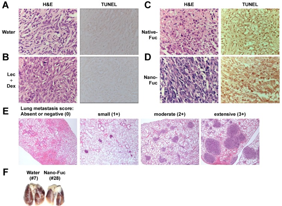
To evaluate the mechanisms of cell death in vivo, analyses were performed using hematoxylin and eosin (H&E) staining and the TUNEL (terminal deoxynucleotidyl transferase-mediated dUTP nick end labeling) assay. Figures 2A and 2B show nuclei stained dark blue with hematoxylin and cytoplasm-stained pink with eosin. In contrast, tumors treated with unprocessed fucoidan or nanoparticle-formulated fucoidan (see Figures 2C and 2D, H&E) exhibited numerous apoptotic cells characterized by nuclear condensation and fragmentation. Concurrently, the density of viable tumor cells was markedly reduced. The TUNEL staining results confirmed that there was a higher level of apoptosis in tumors treated with both unprocessed fucoidan and nanoparticle-formulated fucoidan when compared to the control group, which consisted of LM8-derived tumors (refer to Figures 2C and 2D, TUNEL).
In this model, subcutaneous inoculation of LM8 cells into the backs of C3H mice resulted in the formation of local primary tumors as well as spontaneous metastasis to the lungs. The anti-metastatic effect of fucoidan was confirmed by comparing spontaneous lung metastasis of LM8 cells in tumor-bearing mice treated with fucoidan against a control group treated with water. A four-point scale was used to quantitatively score LM8 cell infiltration into the lungs, with 0 indicating no infiltration, and 1+, 2+, and 3+ representing mild, moderate, and severe infiltration, respectively. The anatomical distribution of metastatic nodules on the lung surface correlated with the microscopic evaluation results (see Figure. 2E, F). Spontaneous lung metastasis was observed in all nine control mice inoculated with LM8 cells, with a mean score of 1.89 ± 0.78.
On the other hand, three of eight mice treated with raw fucoidan or nanoparticle fucoidan in each group showed no lung nodules detectable by macroscopic examination. The mean scores for the raw fucoidan and nanoparticle fucoidan groups were 0.88 ± 0.83 and 1.13 ± 1.13, respectively. Thus, oral administration of raw fucoidan and nanoparticle fucoidan reduced the scores compared to the control group, with a significant difference observed specifically between the raw fucoidan group and the control group. Among mice treated with lecithin and dextrin, spontaneous lung metastasis was observed in five out of six animals, with a mean score of 1.50 ± 1.05. Overall, these findings suggest that both raw fucoidan and nanoparticle fucoidan administration inhibited lung metastasis.
Lastly, transport assays using Caco-2 cells were conducted with unprocessed and nanoparticle-formulated fucoidan. The permeation amount and permeation rate of the nanoparticle-formulated fucoidan were significantly higher than those of the unprocessed fucoidan.
The observation that fucoidan hinders tumor growth in LM8 osteosarcoma and lowers tumor sizes and pulmonary metastases in vivo points to its potential therapeutic application. These results also reveal that the anti-osteosarcoma effects of fucoidan are strengthened by encapsulating it in nanoparticles, in part because this method increases its permeability.
Source: Mar Drugs. 2013 Oct 30;11(11):4267–4278. doi: 10.3390/md11114267