
Vascular occlusion is a condition in which blood flow is blocked by a thrombus, potentially leading to serious health complications. Sulfated polysaccharide-based coatings can be used to prevent “surface-induced thrombosis.”
While natural heparin is one of the most commonly used sulfated polysaccharides acting as an anticoagulant, its long-term use carries a risk of bleeding. In contrast, fucoidan—a marine-derived sulfated polysaccharide—is attracting attention as an alternative anticoagulant that does not carry the risk of bleeding.
In this blog, I would like to inform you of the study, “A New Route of Fucoidan Immobilization on Low Density Polyethylene and Its Blood Compatibility and Anticoagulation Activity” by Kadir Ozaltin et al., which investigates the effects of plasma treatment on LDPE (low-density polyethylene) and the subsequent immobilization of the anticoagulant substances heparin and fucoidan.
First, in the study, low-density polyethylene (LDPE) was used as a base material, and surface mohedification was performed via plasma treatment and graft polymerization. Specifically, polymer brushes derived from allylamine (AAM), N-allylmethylamine (MAAM), and N,N-dimethylallylamine (DAAM)—which possess an amine functional group alongside one or two methyl groups, respectively—were introduced; the study investigated which of these polymer brushes was optimal for the immobilization of the anticoagulant’s heparin and fucoidan.
Samples modified with AAM, MAAM, and DAAM all exhibited similar (and insufficient) anticoagulant properties. Although surface analysis confirmed the formation of these polymer brushes, no significant effect was observed regarding the subsequent immobilization of heparin or fucoidan.
Surface wettability was evaluated by measuring the static water contact angle using the sessile drop technique with distilled water as the test liquid; the results are presented in Table 1. Changes in the water contact angle (θw)—reflecting how water spreads across the surface due to intermolecular interactions—serve as an indicator of hydrophilicity and correlate with surface energy. For the reference polyethylene (PE) material, the water contact angle decreased significantly by approximately 33% (from 85.3° to 56.9°) following plasma treatment. This reduction is attributed to the plasma-induced formation of hydrophilic oxygen-containing functional groups [40] and the increase in surface roughness described in Section 2.3. This enhancement in wettability and surface energy places the PET surface in a state conducive to interactions during subsequent modification processes. Furthermore, such hydrophilic surfaces are known to significantly influence interactions with cells and plasma proteins.
Due to insufficient immobilization of the anticoagulants, the heparin- and fucoidan-immobilized samples before plasma treatment (PEH and PEF, respectively) and the heparin-immobilized sample after plasma treatment (PETH) exhibited nearly identical wettability. In contrast, the fucoidan-immobilized sample after plasma treatment (PETF) showed the lowest water contact angle among all the anticoagulant-immobilized samples, a result attributed to the sample’s hydrophilicity. These findings indicate that fucoidan was successfully immobilized on the surface, a conclusion further confirmed by scanning electron microscopy observations.
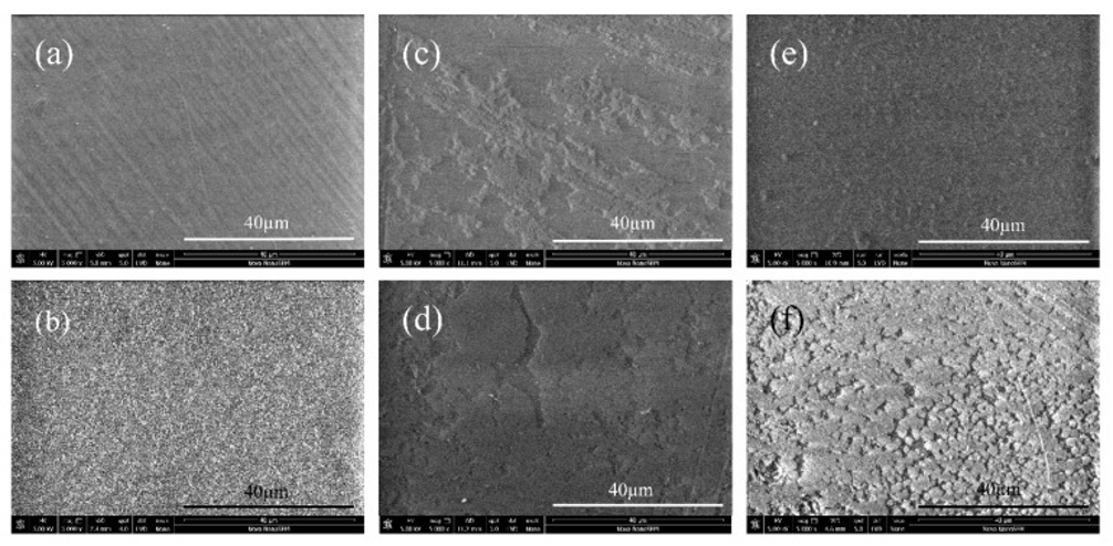
Figure 1 shows SEM images of the surface morphology of the samples. Although the untreated PE sample exhibits a fine, uniform fibrous structure derived from the manufacturing process, it presents a generally homogeneous and relatively smooth surface morphology (Fig. 1a). Upon plasma treatment, the surface morphology became rougher due to surface restructuring caused by plasma exposure (Fig. 1b). Consequently, the surface area increased alongside the surface roughness—a surface state considered desirable for subsequent interactions. Without plasma treatment, the immobilization of the anticoagulant on the PE surface resulted in incomplete adhesion and limited coverage (Figs. 1c, d). As a result, both the immobilized anticoagulant and the substrate itself came into contact with blood, leading to reduced antithrombotic efficacy. In contrast, for the plasma-treated samples (Figs. 1e, f), the functional groups introduced by the plasma treatment led to a more homogeneous, fully continuous coating layer.
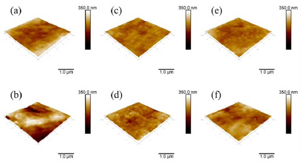
The surface morphology of the samples was examined using AFM, and the resulting images are shown in Figure 2. The surface of the reference PE sample was relatively smooth, with an average roughness of 24.2 nm. When the anticoagulants heparin and fucoidan were directly immobilized onto the PE surface, the surface roughness decreased to 13.9 nm and 20.3 nm, respectively (Figures 2c and 2d). This suggests that heparin and fucoidan particles partially covered the PE pores in a non-uniform manner. In contrast, the PE surface exhibited increased roughness following plasma etching (Figure 2b). A similar trend was observed for the PETH and PETF samples after the immobilization of heparin and fucoidan; the anticoagulant layers reduced the roughness of the PET surfaces to 17.9 nm and 24 nm, respectively. The roughness values for PETH and PETF were slightly higher than those for the corresponding PEH and PEF samples, an effect attributed to the plasma treatment. These results indicate that the roughness value itself is not a direct indicator of coating layer uniformity; the morphological characteristics of the immobilized anticoagulants must also be considered. Furthermore, although the reference PE and PETF samples exhibited similar surface roughness values, their wettability differed significantly; this clearly demonstrates that wettability depends on the chemical properties of the substrate, even when roughness is comparable. Roughness is a crucial property regarding the contact area available for the adhesion of cells and blood proteins.
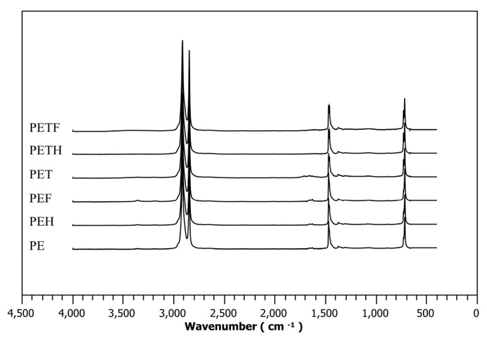
Figure 3 shows the chemical changes near the surface of selected samples, as determined by Attenuated Total Reflection (ATR) Fourier Transform Infrared (FTIR) spectroscopy. In the PE spectrum, major peaks attributable to C–H stretching vibrations are observed at 2915 and 2850 cm⁻¹; additionally, characteristic signals arising from C–H bending vibrations and methylene group rocking vibrations appear at 1470 and 719 cm⁻¹, respectively. For the PET sample, in addition to the peaks derived from PE, peaks attributable to amino groups (introduced via plasma treatment) at 1630 cm⁻¹ and oxygen-containing hydroperoxides at 3370 cm⁻¹ are clearly identified. No significant changes were observed in the PEH and PEF spectra compared to the PET spectrum; this indicates that the immobilization of heparin and fucoidan onto the PE surface is limited in the absence of plasma treatment. Conversely, for the PETH and PETF samples, the peaks associated with hydroperoxides, and amino groups are no longer detectable, indicating that heparin and fucoidan were successfully immobilized on the PE surface following plasma treatment.
For the untreated samples (PE, PEH, PEF), the XPS spectra showed comparable levels of carbon, oxygen, nitrogen, and sulfur, regardless of exposure to the anticoagulant. The constant carbon content suggests that the immobilization of heparin or fucoidan onto the PE surface either did not occur or was limited in extent. As expected, the oxygen content also remained constant, indicating that no oxide groups had been introduced via plasma treatment. Furthermore, the absence of detected nitrogen serves as evidence of the presence of free amino groups.
Following plasma treatment, the carbon content decreased by approximately 12%, while the oxygen content increased significantly due to the introduction of oxide groups. Additionally, the detection of 1.1% nitrogen indicates that amino groups were introduced during the plasma treatment.
In the case of anticoagulant immobilization following plasma treatment (PETH and PETF), chemical bonding resulted in a decrease in carbon and nitrogen (amino group) content, while the oxygen content increased due to the oxygen-containing functional groups present in heparin and fucoidan. In addition to the consumption of carbon and nitrogen, pH values serve as the most critical evidence of anticoagulant immobilization. Based on the pH values, the fucoidan layer immobilized on the PET sample is thicker than the immobilized heparin layer; consequently, it exerts a greater influence on antithrombotic properties.
All coagulation pathways were evaluated using prothrombin time (PT), activated partial thromboplastin time (aPTT), and thrombin time (TT).
PT measurement assesses the time required for clot formation via the extrinsic and common pathways. The normal range for healthy donors is generally 11–13.5 seconds; all samples fell within this range.
aPTT measurement relates to the intrinsic and common pathways. The normal aPTT range for healthy donors is 25–32 seconds. No prolongation of aPTT was observed in any sample, indicating no anticoagulant effect on the intrinsic pathway; this implies the absence of significant inhibitory activity against intrinsic coagulation factors.
The TT value of the control PE (15.9 seconds) was comparable to that of the plasma-treated sample (PET). This indicates that the oxidized functional groups and increased hydrophilicity introduced by plasma treatment did not affect TT. Although hydrophilicity is generally known to influence the coagulation cascade, it is important to note that pH and surface charge are also critical parameters affecting the process. Similar results were observed for samples grafted with AAM, MAAM, and DAAM (PETA, PETM, and PETD, respectively) and for those with immobilized anticoagulants. These findings are consistent with SEM observations. As shown in Figure 4, when the anticoagulants heparin or fucoidan were immobilized without plasma treatment (PEH and PEF), no effect on TT was observed due to insufficient immobilization. This aligns with XPS results indicating that the sulfur components required to enhance antithrombotic activity via heparin cofactor II (HCII) were absent in the PEH and PEF samples. Since sulfur was detected only in the samples with immobilized heparin or fucoidan (PETH and PETF) following plasma treatment, the expected antithrombotic activity was confirmed solely for PETF, which exhibited a TT value of 20.9 seconds. The primary reason for this is that the sulfur content was 2.4% for PETF, whereas it was only 0.2% for PETH. Furthermore, as shown in Figure 4, in contrast to the heparin-immobilized PETH samples, the immobilization of fucoidan on the PET surface was sufficiently uniform, resulting in enhanced anticoagulant activity. In the case of heparin, however, immobilization was achieved via covalent bonding, which prevented sufficient interaction with coagulation factors. For these results, samples with immobilized fucoidan exhibited anticoagulant activity; this was supported by the high uniformity of the immobilization and the fact that fucoidan demonstrated a stronger anticoagulant effect than heparin. Therefore, surface modification of LDPE via plasma treatment followed by fucoidan immobilization is an effective method for preventing surface-induced thrombus formation in blood-contacting biomaterials.
Source: Int J Mol Sci. 2016 Jun 9;17(6):908. doi: 10.3390/ijms17060908