
Helicobacter pylori infection is a common cause of gastritis, gastric ulcers, and ultimately, gastric cancer. The standard medical approach for this condition involves antibiotics, yet the bacteria’s resistance to multiple antibiotics increases the risk of treatment failure in treating Helicobacter pylori.
Fucoidan demonstrates powerful anti-inflammatory properties, making it an effective agent in reducing inflammation. This blog post will discuss the study “Fucoidan from Sargassum hemiphyllum inhibits infection and inflammation of Helicobacter pylori” by Bo-Rui Chen et al. Their research demonstrated that fucoidan extracted from Sargassum effectively lowers Helicobacter pylori infections without contributing to drug resistance.
The objective of this research was to evaluate fucoidan’s toxicity in RAW264.7 macrophage cells and ascertain the level at which no evident cytotoxicity occurred. In other words, no cytotoxicity was observed at fucoidan concentrations up to 1000 μg/mL. To investigate fucoidan’s effect on NO inhibition, we added lipopolysaccharides (LPS) to RAW264.7 cells to induce NO production and set NO production in the LPS-treated group as 100%. In contrast, we measured LPS-induced NO production in RAW264.7 cells treated with various concentrations of fucoidan. The results showed that NO concentration was inversely proportional to the amount of fucoidan added to the cells in a dose-dependent manner. Intracellular NO production showed a significant difference at a fucoidan concentration of 250 μg/mL compared to the LPS group.
Following this, their research focused on fucoidan’s inhibitory effects on H. pylori. The infection ratio of H. pylori to cells was set at 100:1 (multiplicity of infection, MOI = 100). Three test groups were included in the study: post-treatment, pre-treatment, and co-treatment. Bacterial challenge experiments showed that fucoidan was generally effective against H. pylori infection in a dose-dependent manner in the post-treatment group. At a fucoidan concentration of 250 μg/mL, the infection rate was significantly reduced compared to the untreated group. At a fucoidan concentration of 2000 μg/mL, the infection rate was further reduced by 40% in the post-treatment group, but no inhibitory activity was observed in the other two groups. This indicates a direct interaction between fucoidan and H. pylori.
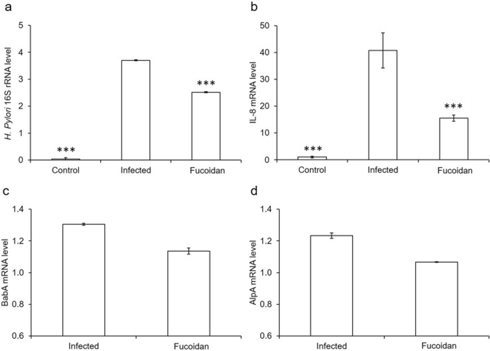
The researchers proposed that fucoidan’s efficacy stemmed from a reduction in H. pylori’s adherence to host cells. Therefore, we harvested H. pylori-infected AGS cells and performed RT-PCR analysis to investigate the mechanism behind this phenomenon. The analysis showed that 16S rRNA expression was significantly reduced in fucoidan-treated cells, as shown in Figure 1a. Fucoidan treatment likely accounts for the diminished IL-8 release from bacterially infected AGS cells, as H. pylori infection typically triggers the secretion of the proinflammatory cytokine IL-8, as illustrated in Figure 1b.
The H. pylori outer membrane proteins (OMPs) AlpA and BabA have been characterized as adhesins or adhesion-associated proteins. Therefore, the researchers analyzed the involvement of these factors in H. pylori infection of AGS cells. The results, as depicted in the figure, confirm that fucoidan exhibits an inhibitory influence on the expression of AlpA and BabA within H. pylori. 1c, d. Therefore, it was concluded that fucoidan is a substance that reduces the ability of H. pylori to adhere to host cells and inhibits the induction of H. pylori-associated inflammatory responses in host cells.
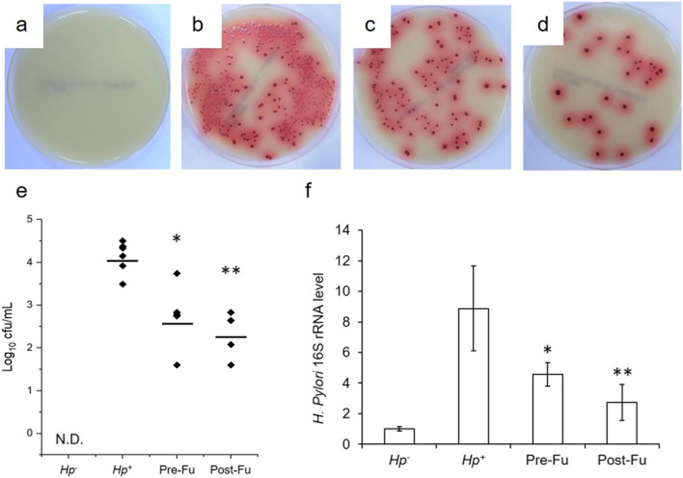
Next, they further evaluated the therapeutic effects of fucoidan on H. pylori-infected mice. Initial assays showed that H. pylori primarily resided in the stomachs of infected mice, consistent with our experimental results. Regarding H. pylori counts, both the pre- and post-treatment groups (Pre-Fu and Post-Fu groups) had significantly lower counts than the Hp+ group, while the Post-Fu group performed relatively better than the Pre-Fu group. H. pylori 16S rRNA is commonly used as an indicator of its expression, and the Hp+ group had the highest 16S rRNA expression level of the three groups. The post-Fu group also demonstrated a greater reduction in the overall H. pylori count in mice compared to the Pre-Fu group, as depicted in Figure 2.
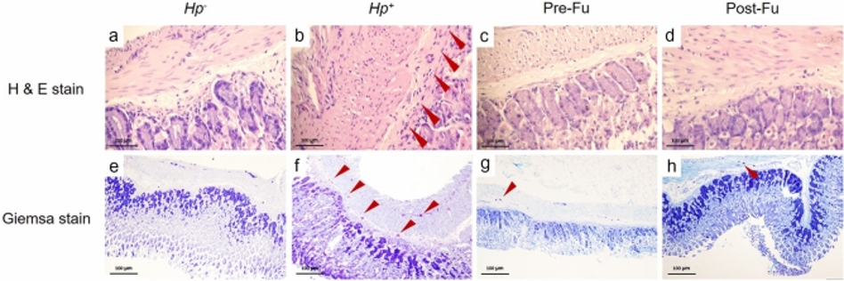
Figure 3 illustrates the application of the Sydney classification system for assessing PMN infiltration, chronic inflammation, and hyperplasia within gastric tissue samples from H. pylori-infected mice. H&E staining revealed significant cellular infiltration in the muscularis mucosae of the infected group, as evidenced by an irregular gastric mucosal layer containing a significant number of immune cells. In contrast, the pathological damage caused by H. pylori was significantly improved in the fucoidan-treated group based on histopathological scores. In the Post-Fu group, there was a decrease in polymorphonuclear leukocyte (PMN) activity, chronic inflammation, and hyperplasia scores relative to the Hp+ group. Consequently, fucoidan was found to be able to modulate the host immune response by ameliorating some adverse symptoms, such as eosinophil infiltration, during H. pylori infection.
Th2 cells are characterized by their production of the cytokines IL-4 and IL-5. To understand whether fucoidan modulates the immune response of H. pylori-infected mice along the Th2 pathway, we measured the expression levels of IL-4 and IL-5. The study found that IL-4 expression levels were not significantly different between the test groups. The IL-5 expression levels, demonstrating a positive correlation with eosinophil infiltration severity, were substantially lowered in the experimental groups. Meanwhile, IL-6, IL-1β, and TNF-α are pro-inflammatory cytokines. Cytokine analysis of IL-6, IL-1β, and TNF-α revealed dramatic increases in BALB/c mice infected with H. pylori for 2 weeks. In contrast, IL-6 and TNF-α expression levels were significantly reduced in infected mice treated with fucoidan. Only IL-1β was significantly reduced in the Post-Fu group.
The results indicate that fucoidan blocks H. pylori adherence to host cells, leading to a more than twofold decrease in infection rates after treatment, notably in the AGS cell model. Fucoidan also inhibited H. pylori BabA and AlpA adhesion and significantly reduced the total number of H. pylori bacteria and the concentrations of IL-6 and TNF-α in vivo. Fucoidan is an effective compound for protecting the stomach from H. pylori, as evidenced by its ability to reduce the bacteria’s numbers and the body’s inflammatory responses.
Source: Sci Rep. 2022 Jan 10;12:429. doi: 10.1038/s41598-021-04151-5