
Peritoneal dissemination, which is the most prevalent form of metastasis observed in particularly aggressive types of gastric cancer, signifies a condition that cannot be surgically removed and for which no successful therapeutic strategies have been developed to date. Studies have revealed that Galectin-4 plays a crucial role in the peritoneal metastasis of gastric cancer cells.
This blog post will present the study “Inhibitory Effect of Fucoidan Analogs on Highly Metastatic Gastric Cancer Cells via Galectin-4 Inhibition” by Shuting Ji et al.
The research operated under the assumption that blocking Galectin-4’s activity would successfully inhibit peritoneal metastasis.
Furthermore, research indicates that fucoidan sourced from Fucus vesiculosus possesses robust inhibitory capabilities when it comes to Galectin-4. The glycan structures and molecular weights of natural fucoidans vary depending on the species of algae from which they are derived; furthermore, differences in extraction methods can introduce variability in both molecular weight and structure. The intricate and dissimilar structures, along with varying sulfation in natural fucoidans, make it a complex challenge to fully understand how these structural features influence their interaction with Galectin-4. However, synthetic fucoidan analogs are characterized by homogenous and distinct structures and sulfation patterns.
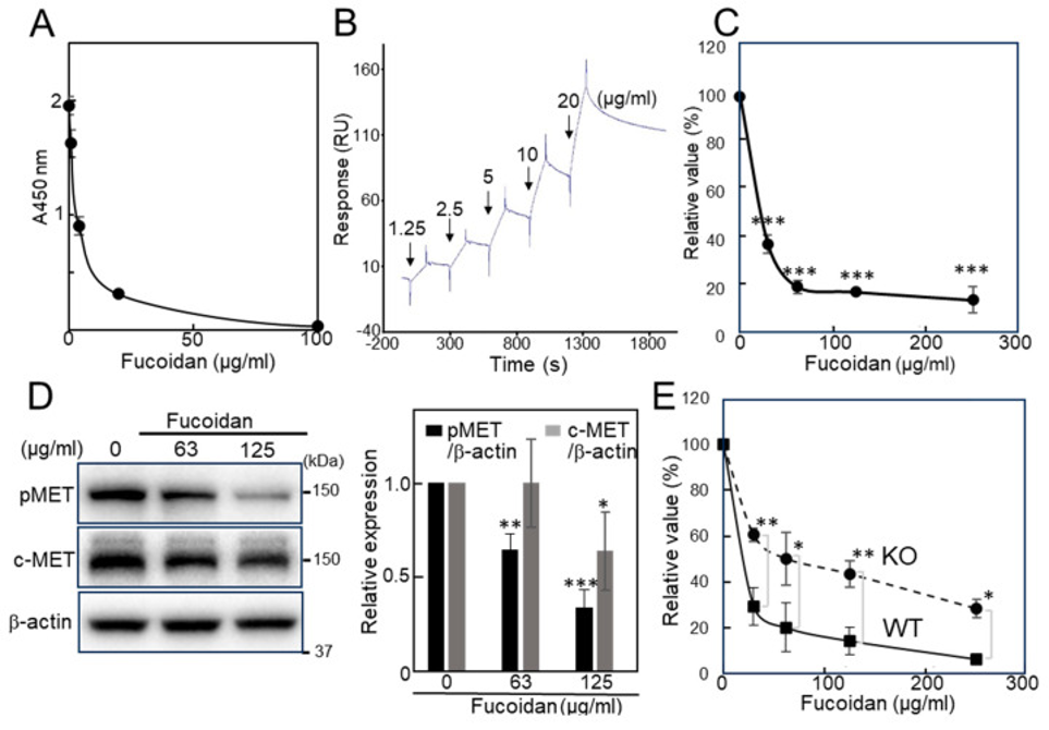
Initially, a competitive enzyme-linked immunosorbent assay (ELISA) was employed to assess the inhibitory effects on Galectin-4 binding. First, the inhibitory activity of natural fucoidan derived from Fucus vesiculosus against Galectin-4 was measured using cholesterol-3-sulfate-coated ELISA plates. The fucoidan inhibited Galectin-4 binding in a dose-dependent manner (see Figure 1A). In addition to the aforementioned steps, Galectin-4 was strategically immobilized onto a sensor chip, enabling the measurement of the fucoidan’s binding affinity for Galectin-4 through the utilization of surface plasmon resonance (SPR) technology, which is depicted in Figure 1B.
Next, the effects of the fucoidan on the proliferation of MKN45 cells were investigated. MKN45 cells are a widely used human gastric cancer cell line derived from a poorly differentiated adenocarcinoma that expresses Galectin-4; an adenosine triphosphate (ATP)-based cell viability assay was employed to assess cell proliferation. The natural fucoidan inhibited the proliferation of MKN45 cells in a concentration-dependent manner, as shown in Figure 1C. Anticipating that the fucoidan might exert effects similar to those observed upon the suppression of Galectin-4 expression, we found that the natural fucoidan indeed demonstrated effects comparable to those of Galectin-4 suppression, inhibiting the expression of both c-MET and pMET (see Figure 1D).
The proliferation of NUGC4 cells, a gastric cancer cell line that is poorly differentiated and notable for its significant expression of Galectin-4 [2], was effectively suppressed by natural fucoidan, much like the observed effects on MKN45 cells, and this suppression showed a clear dependence on the concentration of fucoidan administered (illustrated in Figure 1E). Furthermore, we investigated whether the knockout (KO) of Galectin-4 would influence the inhibitory effects exerted by natural fucoidan. The inhibitory activity of fucoidan was attenuated in the KO cells compared to the wild-type (WT) cells; this observation suggests that Galectin-4 plays a role in the mechanism by which fucoidan exerts its inhibitory effects (see Figure 1E). A Galectin-4-mediated pathway is suggested by these results as a mechanism through which natural fucoidan suppresses gastric cancer cell proliferation.
Despite this, the considerable molecular weight and diverse structure of natural fucoidan present a challenge in accurately identifying the precise structural elements responsible for its strong affinity to Galectin-4. Consequently, further experiments were conducted utilizing synthetic fucoidan.
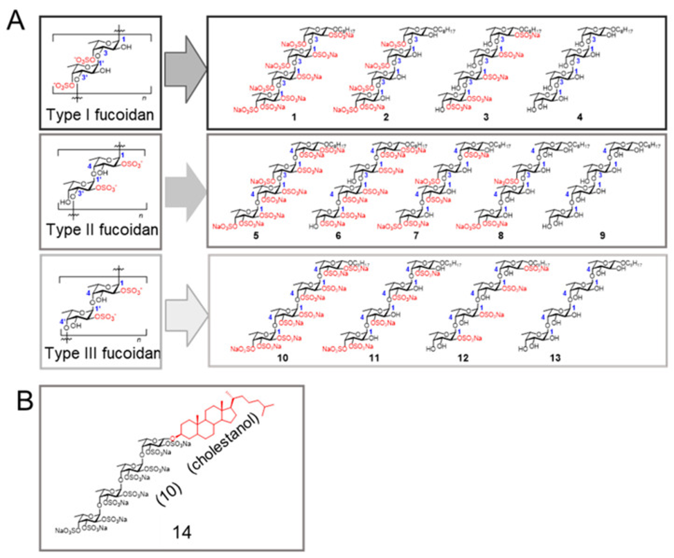
Fucoidans can be classified into three groups based on differences in their sugar backbone chains. The structures of the fucoidan analogs used are shown in Figure 2. Type I, Type II, and Type III fucoidan groups are composed of repeating α(1,3)-linked, α(1,3)- and α(1,4)-linked, and α(1,4)-linked l-fucose, respectively (see Figure 2A). Our strategy was to first select the most potent fucoidan analog from fucoidan analogs 1–13. The fucoidan analog 14, shown in Figure 2B, was subjected to study because it features a cholestanyl group serving as the aglycone moiety, a substitution for the octyl group found in compound 10, and this alteration was hypothesized to potentially amplify inhibitory activity through cholestanol (Cho)-conjugation.
The researchers first evaluated the inhibitory capabilities of all fucoidan analogs (1–14) they created to identify potential candidates. Fucoidan analogs 2, 3, and 14 showed rather stronger inhibitory activities on galectin-4 binding toward cholesterol 3-sulfate and asialofetuin. Next, the inhibitory activity of fucoidan analogs on the proliferation of MKN45 cells was measured. Fucoidan analogs 3 and 10 suppressed cell proliferation, and 14 showed the strongest growth inhibitory activity. On the other hand, the presence of Cho (100 μM) did not elicit a significant inhibitory effect. The inhibitory activities of fucoidan analogs 11 and 12 were comparable; they selected 12 as the control because it has the same sulfation pattern (2-O-sulfated) as the fucoidan analog 3, and natural fucoidan extracted from F. vesiculosus is mainly 2-O-sulfated. From the viewpoint of growth and binding inhibitory activities, we narrowed it down to 2, 3, 10, 12, and 14 for further investigation.
Fucoidan analogs inhibited the proliferative ability of NUGC4 cells, but minimal inhibitory activity was observed against KO cells. 14 showed strong growth inhibitory activity against NUGC4 cells at a concentration of 50 μM, and the inhibitory activity was attenuated against KO cells in the lower concentration range. Galectin-4 appears to be at least partly involved in the growth inhibition observed with fucoidan analogs, based on these findings.
Furthermore, they quantified the extent to which fucoidan analogs suppressed HEK293 cell proliferation. Little inhibitory activity was observed at the tested concentrations, suggesting that the inhibitory activity against gastric cancer cells was not due to simple cytotoxicity.
Blood group A type 1 glycans exhibit a strong affinity for both galectin-4 and SARS-CoV-2. They measured the inhibitory activity of the fucoidan analogs against galectin-4 using blood group A type 1 glycan as a ligand and found that 3, 10, 12, and 14 effectively inhibited the galectin-4 binding to the ligand in a dose-dependent manner. A correlation was found between the strongest inhibitory activity among the tested fucoidan analogs, specifically in compound 14, and its most potent growth-inhibitory effect on cancer cells.
The expression of c-MET and pMET was investigated to clarify the growth-inhibitory mechanism of fucoidan analogs. Distinct suppression of c-MET and pMET was observed in 3-, 10-, and 14-treated cells in both MKN45 and NUGC4 cells. Real-time polymerase chain reaction was performed to examine whether the reduction in c-MET in fucoidan analog-treated cells occurred at a genetic level. The genetic expression of c-MET remained constant in the cells that underwent treatment, indicating that the decrease was a result of post-translational modifications.
Researchers utilized a surface plasmon resonance (SPR) system, where Galectin-4 was immobilized, to directly measure the affinity of fucoidan analogs for this protein. Some of the fucoidan analogs exhibited a behavior characterized by slow dissociation from the immobilized Galectin-4 on the sensor chip, even after the injection had ceased. Consequently, it was not possible to determine precise Kᴅ values. As a result, the scientists decided to determine the relative binding affinities for Galectin-4 by analyzing the individual sensor grams. Fucoidan analogs 2, 3, 10, 12, and 14 demonstrated dose-dependent binding to Galectin-4. Conversely, fucoidan analogs 4, 9, 13, and Cho did not exhibit any discernible binding response. A noteworthy observation is that analog 14 exhibited a substantially stronger reaction during the binding stage. Based on the obtained data, it is possible to infer that the immobilization of Galectin-4 onto the sensor chip could have played a role in promoting the aggregation of compound 14.
Source: Int J Mol Sci. 2025 Sep 21;26(18):9228. doi: 10.3390/ijms26189228