As the global population continues to age, neurodegenerative diseases are expected to pose a substantial threat to the well-being of people across the world. However, existing therapies are unable to provide a fundamental cure, serving merely to alleviate disease symptoms at best. Conversely, preclinical studies suggest that “oxidative stress, which plays a pivotal role in the neurodegenerative process—could serve as a promising therapeutic target for the development of novel treatments.
In this blog. I would like to share the following study, “The protective effect of non-invasive low-intensity pulsed electric field and fucoidan in preventing oxidative stress-induced motor neuron death via ROCK/Akt pathway” by Chih-Hsiung Hsieh et al.
Fucoidan, a sulfated polysaccharide obtained from brown algae like Fucus vesiculosus, has undergone significant research into its biological functions, including its anticoagulant, anticancer, immunomodulatory, and anti-inflammatory effects. Of particular note, recent studies have demonstrated that fucoidan exerts protective effects against the death of dopaminergic neurons in both in vitro and in vivo models of Parkinson’s disease.
To determine if non-contact, non-invasive low-intensity pulsed electric field (LIPEF), alone or with fucoidan, could protect neuronal cells from H2O2 damage, NSC-34 cells were first pretreated for an hour with either LIPEF by itself or LIPEF in conjunction with fucoidan. Subsequently, while maintaining the LIPEF-alone or LIPEF-plus-fucoidan treatment, the cells were further exposed to 100 μM H2O2 for an additional 24 hours. Following the 24-hour exposure, the viability of the NSC-34 cells declined to 53% of the control group’s value. In contrast, in the cells treated with LIPEF, cell viability recovered, rising to 58%, 67%, 76%, and 71% of the control group’s value, respectively. Furthermore, when various concentrations of fucoidan (ranging from 0 to 200 μg/mL) were combined with the 60 V/cm LIPEF treatment, fucoidan most effectively enhanced the protective effects of LIPEF, resulting in cell viability exceeding 85% of the control group’s value. These results demonstrate that LIPEF treatment alone is capable of protecting NSC-34 cells from oxidative stress. Moreover, a combination of fucoidan and LIPEF demonstrated increased protective efficacy compared to fucoidan alone, which was not significantly protective.
Furthermore, to investigate whether intracellular ROS production is reduced when NSC-34 cells undergo combination therapy, ROS levels were analyzed using a DCFH-DA assay. When cells were exposed to 100 μM H2O2 for 24 hours without treatment with either LIPEF or fucoidan, DCF fluorescence intensity significantly increased to as much as 550% of the control value. When H2O2-challenged cells were treated with either LIPEF alone (at 60 V/cm) or fucoidan alone (at 100 μg/ml), it was found that ROS levels were clearly reduced compared to the control value.
Also, the synergistic effect of LIPEF and fucoidan markedly diminished the ROS levels that H2O2 had raised, relative to the control group. Additionally, given that impaired GSH metabolism is a key characteristic of persistent oxidative stress in motor neuron degeneration, the GSH/GSSG ratio in NSC-34 cells was also measured. When cells were exposed to H2O2 for 24 hours without treatment with either LIPEF or fucoidan, the GSH/GSSG ratio significantly decreased. Of particular note is that the combination therapy of LIPEF and fucoidan was able to prevent or mitigate GSH depletion in the H2O2-treated NSC-34 cells. As shown by these results, the protective impact of this combination therapy is potent in mitigating oxidative stress in NSC-34 cells exposed to H2O2.
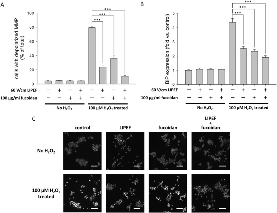
To provide more detail on the connection between mitochondrial membrane potential (MMP) and the protective advantages of LIPEF, used either independently or alongside fucoidan, the researchers evaluated MMP levels using DiOC6(3). As shown in Figure 1A, while the proportion of cells exhibiting MMP depolarization was 4.5% in the control group, this proportion increased dramatically in the group of cells exposed to 100 μM H2O2 for 24 hours without prior treatment with LIPEF or fucoidan. Consequently, it became evident that LIPEF alone can suppress the H2O2-induced loss of MMP. Additionally, the simultaneous application of LIPEF and fucoidan substantially prevented the reduction in mitochondrial membrane potential (MMP) caused by H2O2, consequently lowering the percentage of cells showing MMP depolarization.
The researchers then proceeded to investigate the effect of the combined LIPEF and fucoidan treatment on cellular ER stress. To this end, they measured the expression levels of the BiP protein. As shown in Figure 1B, when NSC-34 cells were exposed to 100 μM H2O2 for 24 hours without prior treatment with LIPEF or fucoidan, BiP expression levels increased dramatically. It was confirmed that treatment with either LIPEF or fucoidan alone could suppress the H2O2-induced increase in BiP expression. Moreover, in the group receiving the combined treatment, the suppressive effect on BiP expression in H2O2-treated cells was further enhanced. Using DAPI staining, the study examined the protective effects of these treatments on the nuclear morphology of NSC-34 cells.
The data presented in Figure 1C demonstrate that exposing NSC-34 cells to 100 μM H2O2 for a duration of 24 hours resulted in a significant induction of nuclear chromatin condensation within these cells. It was confirmed that treatment with either LIPEF or fucoidan alone could suppress H2O2-induced nuclear chromatin condensation. Furthermore, when LIPEF and fucoidan were administered together, they exhibited a notable effect of nearly entirely preventing the nuclear chromatin condensation that is brought about by H2O2, as can be observed in Figure 1C. These results indicated that the protective action of this combination therapy could suppress the apoptotic response in NSC-34 cells exposed to ROS stress.
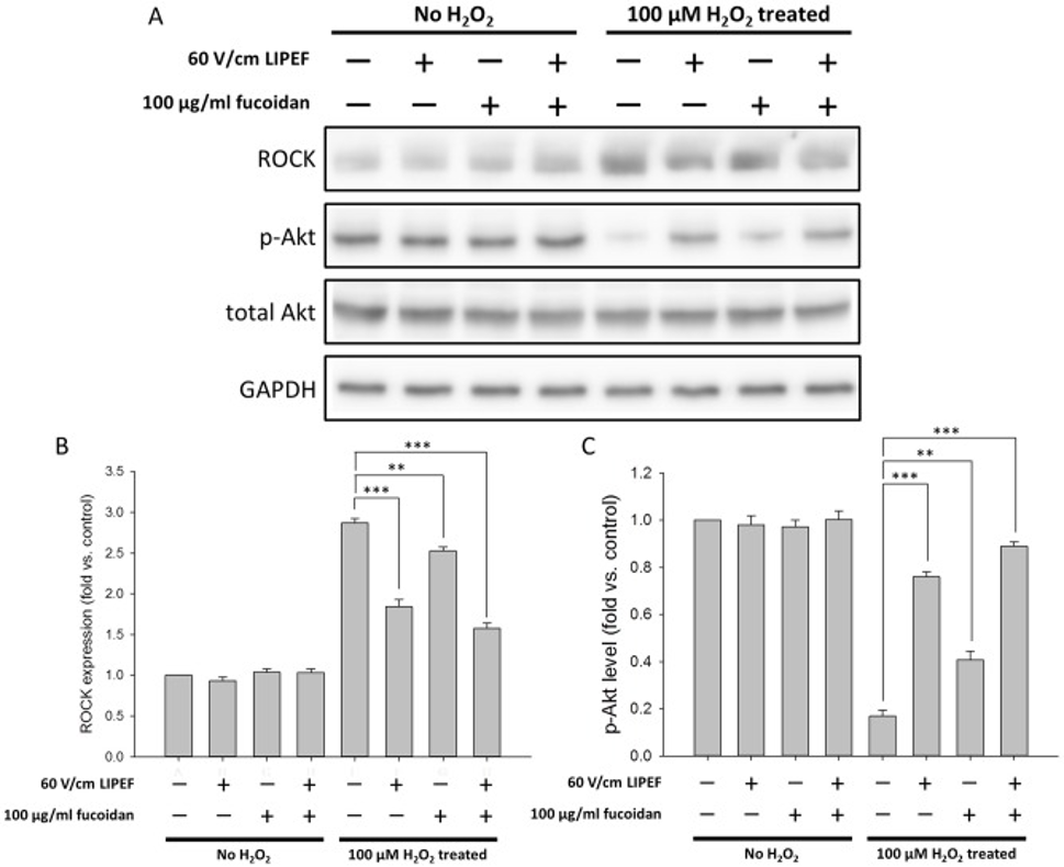
The ROCK pathway has been reported to mitigate oxidative damage in various cell types. Therefore, to further determine whether the protective effects exerted by LIPEF and fucoidan are associated with the ROCK pathway, the researchers examined the protein expression of ROCK in cells that had been pretreated with LIPEF and fucoidan for one hour before a 24-hour exposure to H2O2. As shown in Figure 2A, ROCK expression was significantly increased in cells exposed to 100 μM H2O2 for 24 hours without prior treatment with LIPEF or fucoidan. In Figure 2B, they observed that fucoidan slightly attenuated the H2O2-induced increase in ROCK expression.
In contrast, both treatment with LIPEF alone and the combined treatment with LIPEF and fucoidan could significantly suppress the H2O2-induced increase in ROCK expression. These results demonstrate that the neuroprotective effects conferred by LIPEF are linked to the suppression of ROCK expression, which is upregulated by H2O2. Subsequently, to assess whether Akt also plays a role in these protective effects, the researchers measured the phosphorylation levels of Akt. As shown in Figure 2C, compared to untreated control cells, the levels of phosphorylated Akt (p-Akt) were markedly reduced in cells exposed to 100 μM H2O2 for 24 hours without prior treatment with LIPEF or fucoidan.
The investigation revealed that the application of fucoidan singularly resulted in an insignificant influence on the reduction of Akt phosphorylation caused by H2O2. In both the LIPEF-alone treatment group and the combined LIPEF and fucoidan treatment group, a significant recovery in Akt phosphorylation was observed. These results suggest that LIPEF promotes cell survival by delivering physical stimulation to neurons, thereby activating protective signals via the ROCK pathway while simultaneously reactivating Akt signaling.
Bcl-2 family proteins function as regulators of cell death under conditions of ROS stress. In cells subjected to H2O2-induced stress, combined treatment with LIPEF and fucoidan was found to result in the significant upregulation of Bcl-2 expression and the significant downregulation of Bax expression. It is worth noting that the decrease in the Bax/Bcl-2 ratio, elevated by H2O2, was more pronounced when fucoidan was used alone than when LIPEF was used alone. These results suggest that, within the context of the combined treatment, the regulatory control of Bcl-2 family protein expression plays a role in mediating the protective effects of fucoidan. The evidence collectively suggests that the concurrent application of LIPEF and fucoidan has a synergistic effect in protecting cells from oxidative damage.
To understand the distribution and structure of neuronal cells, this study performed immunofluorescence microscopy, specifically using β-III tubulin staining. When NSC-34 cells were exposed to 100 μM H2O2 for 24 hours without prior treatment with LIPEF or fucoidan, a marked reduction in both the average neurite length and the average number of neurites per cell was clearly observed. Furthermore, treatment with fucoidan alone was found to exert a modest inhibitory effect on the reduction of both neurite length and neurite number. In contrast, treatment with LIPEF alone—as well as combined treatment with LIPEF and fucoidan—significantly suppressed the H2O2-induced degeneration of neurites.
These findings indicate that when NSC-34 cells experience oxidative stress caused by H2O2, applying non-contact LIPEF can help increase the survival rate of neuronal cells and also stop neurite degeneration. What’s more, the findings of this study demonstrated that the simultaneous application of non-contact LIPEF and fucoidan leads to augmented neuroprotection. In summary, these results indicate that the simultaneous application of non-contact LIPEF and fucoidan shows considerable promise for practical use in preventing neurite degeneration and neuronal cell death resulting from oxidative stress.
Source: PLoS One. 2019 Mar 19;14(3):e0214100. doi: 10.1371/journal.pone.0214100