
Cancer-related mortality is frequently attributed to metastasis, which is the process of the disease spreading to other parts of the body. This process is closely connected to various biological functions of cancer cells, such as their growth, spread, attachment, and metastasis through the bloodstream and lymphatic system. Fucoidan derived from the sporophylls of Undaria pinnatifida (Ups-fucoidan) is a subject of interest; however, information regarding the specific effects of Ups-fucoidan on tumor invasion and metastasis remains scarce.
This blog post will therefore introduce a study by Peisheng Wang et al. that examines the “Anti-Metastasis Effect of Fucoidan from Undaria pinnatifida Sporophylls in Mouse Hepatocarcinoma Hca-F Cells.” This research investigated the effects of Ups-fucoidan on cancer cell invasion and metastasis using the Hca-F mouse hepatocarcinoma cell line—a model known to exhibit high invasive potential and lymphatic metastatic capacity in both in vitro and in vivo settings.
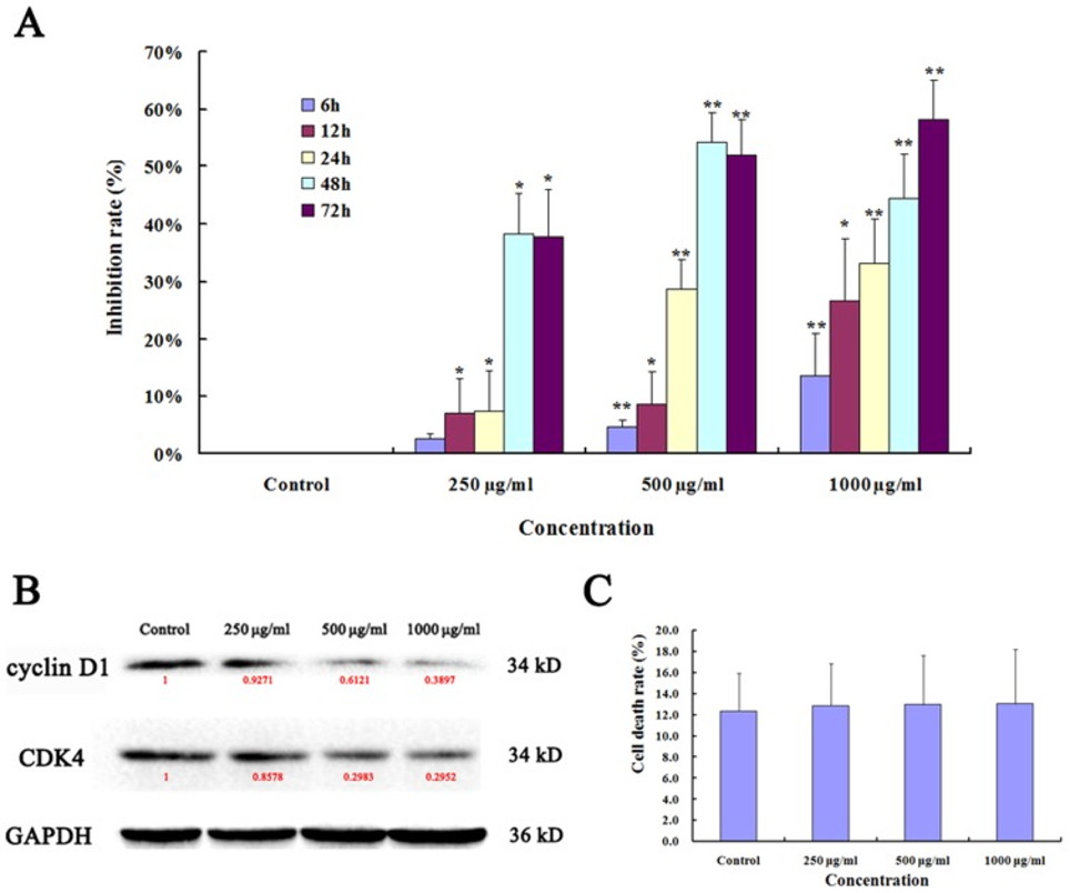
First, the effects of Ups-fucoidan on the proliferation of Hca-F cells were evaluated in vitro by measuring cell viability using an MTT assay. Following treatment with various concentrations of Ups-fucoidan, cell proliferation rates showed statistically significant differences compared with the untreated control group (see Figure 1A). Western blot analysis revealed that Ups-fucoidan reduced the expression levels of both Cyclin D1 and CDK4 (see Figure 1B). After removing the Ups-fucoidan, the number of dead cells was quantified by Trypan blue staining to calculate the cell death rate (D%); however, no statistically significant differences in D% were observed among the Ups-fucoidan concentration groups compared to the control group. The findings indicate that despite Ups-fucoidan’s capacity to impede the proliferation of Hca-F cells, it does not demonstrate any cytotoxic effects, meaning it does not actively kill cells.
The binding capacity of Hca-F cells for peripheral lymphatic endothelial cells was measured using an adhesion assay that followed. The adhesion capacity of Hca-F cells treated with 500 µg/ml and 1000 µg/ml of Ups-fucoidan was significantly lower compared to that of the control group. The adhesion molecule L-selectin plays a crucial role in the adhesion of tumor cells to lymph nodes. L-selectin expression is also observed in tumor cells exhibiting a high potential for lymphatic metastasis. These results suggest that Ups-fucoidan suppresses the expression of L-selectin, thereby inhibiting the adhesion of Hca-F cells to peripheral lymphatic endothelial cells.
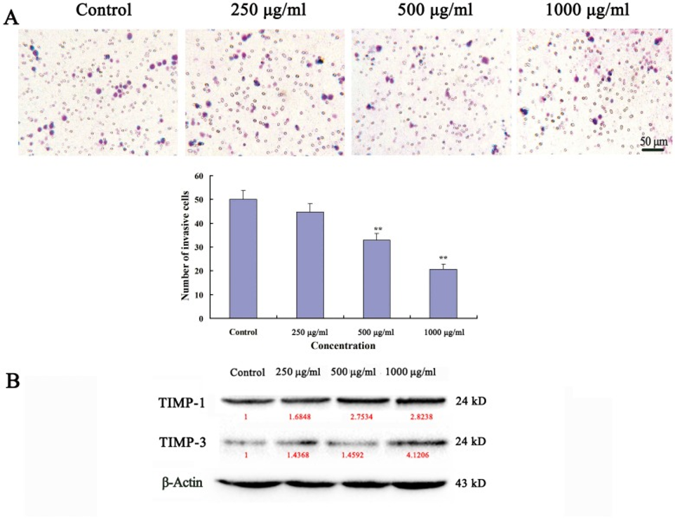
During the invasion assay, it was observed that Ups-fucoidan effectively suppressed the in vitro invasion of Hca-F cells. The number of Ups-fucoidan-treated cells that invaded through Matrigel-coated filters was significantly lower compared to the control group (see Figure 2A). Furthermore, Western blotting analysis revealed that the relative expression levels of TIMP-1 and TIMP-3 were increased compared to the control group (see Figure 2B).
The invasion assay demonstrated that Ups-fucoidan’s activity inhibited Hca-F cells’ in vitro invasion. The number of Ups-fucoidan-treated cells that invaded through Matrigel-coated filters was significantly lower compared to the control group. Furthermore, Western blotting analysis revealed that the relative expression levels of TIMP-1 and TIMP-3 were increased compared to the control group.
Western blotting and ELISA were employed to assess the activity of NF-κB-dependent PI3K/Akt and ERK signaling pathways, as well as the expression of associated key factors. In Ups-fucoidan-treated cells, a reduction in the expression of VEGFR-3, c-MET, p-PI3K, p-Akt, p-ERK1/2, and NF-κB was observed. Furthermore, ELISA results demonstrated that the levels of VEGF-C and HGF proteins in Ups-fucoidan-treated cells were significantly lower compared to the control group (p < 0.01).
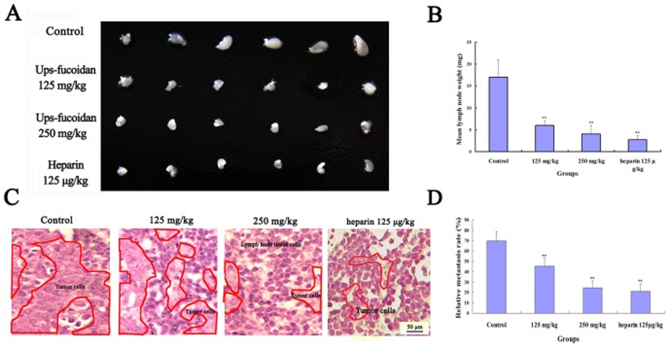
In a study with 615 mice, the group that received Ups-fucoidan after inoculation with Hca-F cells exhibited a notable reduction in the mean weight of lymph node metastases when contrasted with the control group given physiological saline (NS). Figures 3A and 3B illustrate the appearance of the axillary lymph nodes in the Ups-fucoidan-treated group. The mean weight of the axillary lymph nodes in the Ups-fucoidan-treated group was also significantly lower compared to that of the control group. Heparin, which was used as a positive control, demonstrated a clear effect in suppressing lymph node metastasis. At the same time, it triggered a pronounced inclination to hemorrhage. Histological analysis revealed that Ups-fucoidan inhibited the infiltration of Hca-F cells into the lymph nodes (see Figures 3C and 3D). These results suggest that Ups-fucoidan is capable of suppressing tumor metastasis to peripheral lymph nodes in vivo.
The findings indicate that Ups-fucoidan impedes the multiplication, adherence, invasion, and metastatic capabilities of Hca-F cells. This inhibition appears to be achieved through pathways that deactivate the NF-κB signaling cascade, specifically by influencing the PI3K/Akt and ERK signaling routes.
Source: PLoS One. 2014 Aug 27;9(8):e106071. doi: 10.1371/journal.pone.0106071