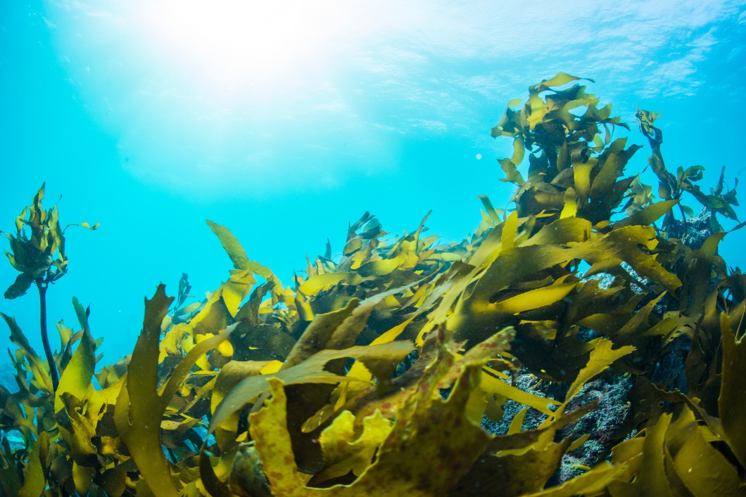
Pulmonary fibrosis is a condition marked by the lungs becoming inflamed and progressively scarred. The principal causes of this condition include smoking, long-term exposure to detrimental agents such as dust, infections, radiation, and specific medications. It is a chronic, progressive, and ultimately fatal form of interstitial lung disease. Currently, no successful treatments exist to enhance the outlook or reverse the progression for individuals suffering from this ailment.
This blog post will introduce you to the study “Isolation, Characterization, and Anti-Idiopathic Pulmonary Fibrosis Activity of a Fucoidan from Costaria costata” by Sijie Wei and colleagues. Researchers isolated fucoidan from Costaria costata, a brown alga, and investigated its inhibitory effects on idiopathic fibrosis using laboratory-based and whole-organism models.
Fucoidan derived from Costaria costata has been reported to possess a wide range of biological activities, including anti-hyperlipidemic effects, inhibitory effects on skin photoaging, and protective effects against carbon tetrachloride (CCl4)-induced liver injury.
Chemical composition analysis of the ethanol precipitate revealed that C. costata polysaccharide (CCP) contains galactose and fucose as its major monosaccharides and has a sulfate group content of 18.54%.
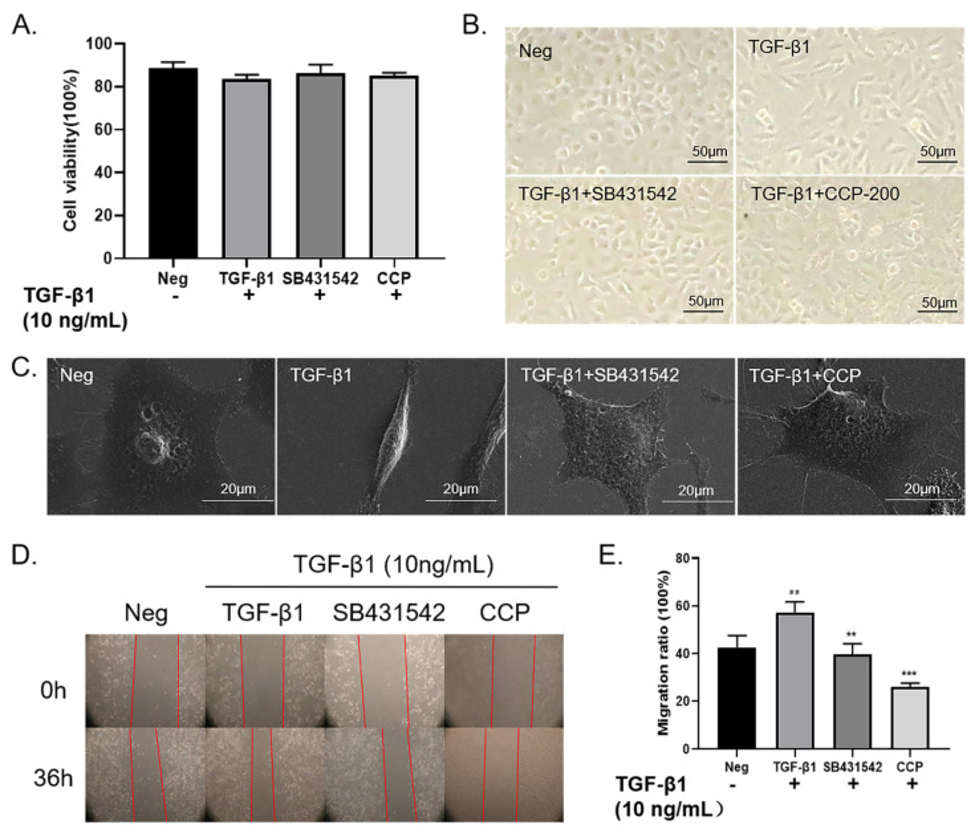
EMT (Epithelial-Mesenchymal Transition) is a biological process in which epithelial cells lose their cellular polarity and cell-cell adhesion functions, transforming into mesenchymal cells that possess migratory and invasive capabilities. TGF-β1 plays a pivotal role in this EMT process, serving as an inducer that drives the phenotypic conversion of polygonal epithelial cells into spindle-shaped fibroblastic cells. In the study, EMT was induced in A549 cells using 10 ng/mL of TGF-β1; subsequently, the cells were treated with either CCP (200 μg/mL) or SB431542 (4 μM) and cultured for 36 hours. Following this treatment, a cytotoxicity assay using the MTT method was performed on the A549 cells. The results demonstrated that no significant changes in cell viability were observed in any of the treatment groups: the TGF-β1-alone group, the TGF-β1 + SB431542 group, or the TGF-β1 + CCP group (see Figure 1A).
As illustrated in Figure 1B, the morphology of the cells induced by TGF-β1—when compared to that of the negative control (normal A549 cells), which exhibited a polygonal, epithelial-like appearance—showed diminished cell-cell adhesion and a transformation into an elongated, spindle-shaped morphology. From the perspective of cellular morphology, TGF-β1 induction not only shifted the cellular phenotype from the normal polygonal, epithelial-like form to a typical elongated, spindle-shaped form but also exerted an effect that attenuated cell-cell adhesion. In comparison to the TGF-β1-induced group, the cell groups treated with 4 μM of SB431542 or 200 μg/mL of CCP exhibited a restoration of cell-cell adhesion and a recapitulation of the normal polygonal epithelial cell morphology.
TGF-β1 influences cell migration and dedifferentiation, and it also controls the expression of extracellular matrix (ECM) components like fibronectin, elastin, and collagen. They used a wound healing assay to determine if CCP could prevent EMT by evaluating the cells’ ability to migrate and invade (as shown in Figures 1D, E). The results demonstrated that cells treated with TGF-β1 exhibited a dramatic enhancement in migration compared to untreated A549 cells. Furthermore, it was confirmed that treatment with either SB431542 or CCP was able to suppress this enhanced migration associated with EMT, albeit to varying degrees. Based on these findings, they have demonstrated—from the perspective of cellular migration—that CCP can inhibit TGF-induced EMT in A549 cells.
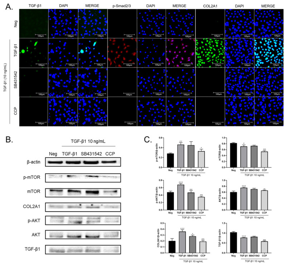
The epithelial-mesenchymal transition (EMT) pathway, when initiated by TGF-β1, is broadly divided into two distinct types: the Smad-dependent and Smad-independent pathways. Analysis using immunofluorescence revealed that the addition of TGF-β1 to A549 cells resulted in a significant increase in the expression levels of TGF-β1, p-Smad2/3, and COL2A1. Figure 2A demonstrates that, by way of contrast, the application of SB431542 or CCP had the effect of diminishing the expression of these specific molecules. These results suggest that CCP may suppress the progression of EMT in lung cancer by acting through the TGF-β/Smad pathway and its downstream molecular mechanisms.
As shown in Figures 2B and 4C, the addition of TGF-β1 significantly upregulated the expression of TGF-β1, COL2A1, mTOR, p-mTOR, AKT, and p-AKT. In contrast, in the group treated with CCP, the increase in protein expression induced by TGF-β1 was significantly suppressed. Meanwhile, regarding the suppressive effects observed in the SB431542-treated group, no statistically significant differences were observed for p-mTOR, mTOR, or AKT. These results are consistent with the findings from the immunofluorescence analysis, suggesting that CCP attenuates TGF-β1-induced EMT in A549 cells by inhibiting both the PI3K/AKT/mTOR pathway and the TGF-β1/Smad pathway.
Generally, the epithelial-mesenchymal transition (EMT) pathway induced by TGF-β1 is broadly categorized into two types: Smad-dependent and Smad-independent pathways; among these, the TGF-β1/Smad signaling pathway has been studied in the greatest detail. Analysis using immunofluorescence revealed that the addition of TGF-β1 to A549 cells resulted in a significant increase in the expression levels of TGF-β1, p-Smad2/3, and COL2A1. Conversely, the administration of SB431542 or CCP led to a reduction in the expression of these molecules (Figure 2A). The results indicate a potential mechanism where CCP inhibits EMT progression in lung cancer by influencing the TGF-β/Smad pathway and its downstream molecular elements.
As shown in Figures 2B and 4C, the addition of TGF-β1 significantly upregulated the expression of TGF-β1, COL2A1, mTOR, p-mTOR, AKT, and p-AKT. In contrast, in the group treated with CCP, the increase in protein expression induced by TGF-β1 was significantly suppressed. Meanwhile, regarding the suppressive effects observed in the SB431542-treated group, no statistically significant differences were observed for p-mTOR, mTOR, or AKT. These results are consistent with the findings from the immunofluorescence analysis, suggesting that CCP attenuates TGF-β1-induced EMT in A549 cells by inhibiting both the PI3K/AKT/mTOR pathway and the TGF-β1/Smad pathway.
As shown in Figures 2B and 2C, induction by TGF-β1 significantly upregulated the expression of TGF-β1, COL2A1, mTOR, p-mTOR, AKT, and p-AKT. The administration of CCP significantly suppressed the increase in protein expression induced by TGF-β1; however, regarding the reductive effects of SB431542 administration, no statistically significant differences were observed for p-mTOR, mTOR, or AKT. These results are consistent with the findings from immunofluorescence analysis, indicating that CCP attenuates TGF-β1-induced EMT in A549 cells by inhibiting the PI3K/AKT/mTOR and TGF-β1/Smad pathways.
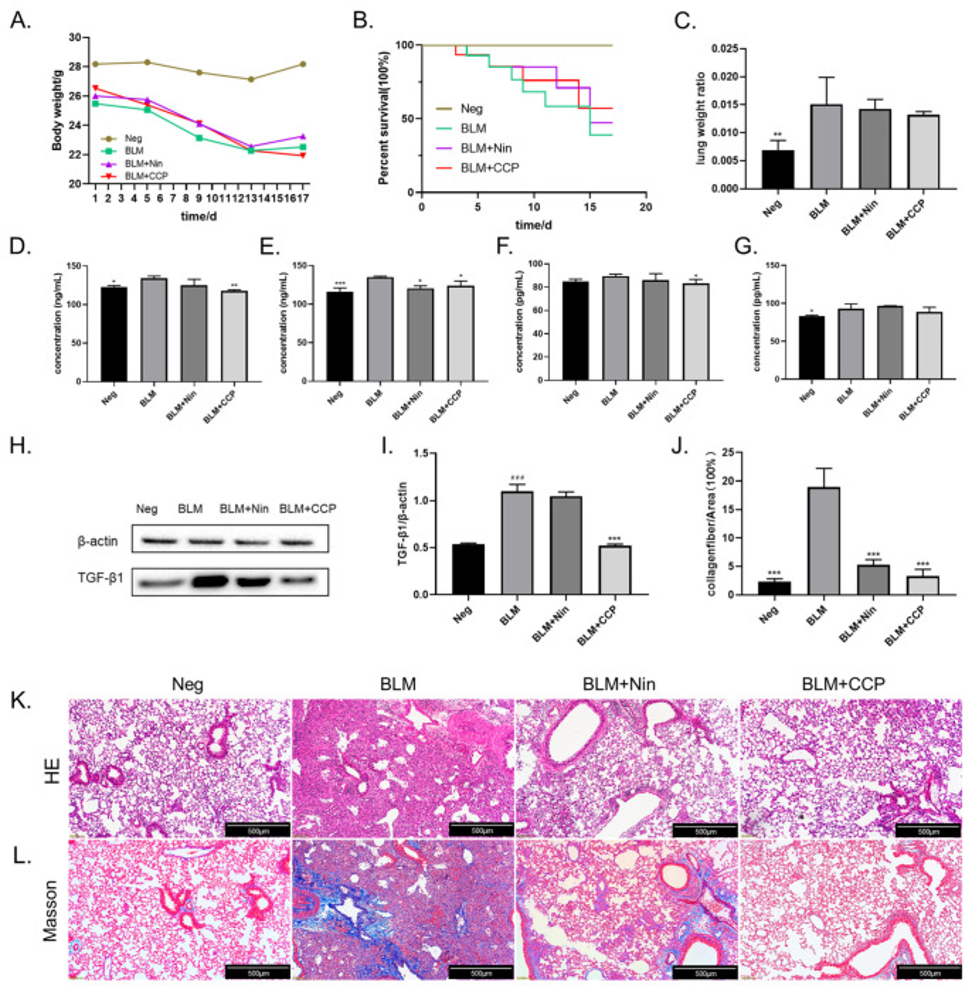
The establishment of a fibrosis model was achieved by employing intratracheal bleomycin (BLM) administration as a method to promptly induce pulmonary fibrosis (PF). The body weight of the mice showed a downward trend from Day 1 to Day 13 following treatment. Among the groups, the BLM-induced group exhibited the most pronounced weight loss, while the CCP-treated group showed the smallest magnitude of reduction. In all groups except for the CCP group, weight loss ceased after Day 13. In the control group, body weight remained at normal levels. These results suggest that while the surgical procedure itself caused some degree of weight loss in the mice, treatment with CCP mitigated this reduction.
In terms of survival statistics, the control group that did not receive any treatment, designated as Neg, exhibited a complete survival rate of 100%. The lowest survival rate among the surgical groups was observed in the BLM-induced group (BLM), with the Nin-treated group (BLM + Nin) showing a slightly better outcome. In contrast, the CCP-treated group exhibited the most favorable survival rate, as shown in Figure 3B. Furthermore, the “lung weight-to-body weight ratio” was calculated for the mice following necropsy. The findings indicated that the lung weight-to-body weight ratio was notably greater in the BLM-induced group compared to the Neg group, probably because of inflammatory reactions and fluid buildup in the lungs. In contrast, a decrease in lung weight was observed in both the Nin-treated and CCP-treated groups (see Figure 3C).
In the lung tissue of patients with Idiopathic Pulmonary Fibrosis (IPF), the overexpression of TGF-β1 is frequently observed as a key characteristic. TGF-β1 levels in the bronchoalveolar lavage fluid of the mice were measured using an ELISA (Fig. 3D). The findings revealed that TGF-β1 levels were higher in the BLM-induced group but decreased in both the Nin-treated and CCP-treated groups when contrasted with the BLM group. Similar results regarding TGF-β1 expression were also obtained from serum samples (Fig. 3E). While the onset of IPF is often accompanied by inflammatory responses, the IL-6–JAK2–STAT3/STAT1 pathway has previously been identified as a key mechanism for the effective treatment of IPF.
The researchers measured IL-6 levels in both bronchoalveolar lavage fluid and serum from mice, demonstrating that CCP significantly reduced IL-6 expression compared to mice treated with BLM (Figures 3F, G). Additionally, the quantified TGF-β1 expression in mouse lung tissue using Western blotting is depicted in Figures 3H, I. The results revealed a significant increase in TGF-β1 expression in the BLM group. Compared to the BLM group, mice treated with Nin showed a trend toward reduced TGF-β1 expression levels; however, this difference did not reach statistical significance. In contrast, treatment with CCP significantly reduced TGF-β1 expression levels in the lung tissue. These findings suggest that the administration of CCP ameliorates pulmonary fibrosis in mice by downregulating TGF-β1 expression.
H&E and Masson staining were performed on lung sections from BLM-treated mice in order to assess the histopathological changes within the lung tissue. Representative images from the H&E staining are presented in Figure 3K. In the negative control group, the structure of the alveolar spaces was well-preserved, and the thickness of the alveolar septa appeared normal. This study clearly demonstrated that BLM induces severe alveolar and interstitial damage—accompanied by inflammation—as well as severe fibrotic lesions in the lungs.
After treatment with CCP and Nin, the lung tissue showed fewer pathological changes. Cellular hyperplasia decreased, and damage to the alveolar regions and the lung’s overall structure was lessened. The results of the Masson staining (see Figure 3L) indicated that BLM administration induced excessive deposition of mature collagen within the mouse lung tissue. Compared to the BLM group, collagen deposition was significantly reduced following treatment with both CCP and Nin. However, a minor amount of residual collagen deposition remained evident in the Nin group. Quantitative analysis of the Masson staining results (see Figure 3J) revealed that, compared to the BLM group, the proportion of collagen deposition decreased by 13.67% and 16.01% in the Nin-treated and CCP-treated groups, respectively.
As a result, it was demonstrated that CCP can prevent the epithelial-mesenchymal transition (EMT) triggered by TGF-β1 in A549 cells by blocking signaling through the TGF-β/Smad and PI3K/AKT/mTOR pathways. Additionally, in vivo research showed that CCP treatment improved bleomycin (BLM)-induced fibrosis and inflammation in mouse lung tissue. The combined data suggest that CCP could protect lung tissue from fibrosis by suppressing and mitigating EMT and inflammation in lung cells.
Source: Molecules. 2023 May 25;28(11):4343. doi: 10.3390/molecules28114343