
Radioactive iodine (RI) can damage the cells within the salivary glands (SGs), preventing them from working properly. This blog post presents the research of Young-Mo Kime et al., titled “Fucoidan attenuates radioiodine-induced salivary gland dysfunction in mice,” which investigates fucoidan’s potential to mitigate RI-induced salivary gland issues in mice.
Alternatively, fucoidan, which is a sulfated polysaccharide that is present in brown algae, exhibits a number of biological activities, such as antioxidant, anti-inflammatory, and immunomodulatory effects.
It was determined that before the experiment, the groups did not have any considerable differences in their body weight. However, after the experiment, the mice in the RI group weighed significantly less than the mice in the normal control group and the mice in the fucoidan group. At 2 and 12 weeks after administration, the mice in the RI group weighed significantly more than the mice in the RI group. When assessing gland weights at 2, 4, and 12 weeks post-RI, the RI group showed a tendency towards lower weights in comparison to the normal and fucoidan groups; however, these observed differences did not reach a level of statistical significance.
The study revealed notable distinctions in the lag time between the groups, specifically at two weeks, four weeks, and twelve weeks after the RI administration. The lag time in the normal group was shorter than that of the RI group. At 4 and 12 weeks after RI, the lag time in the fucoidan-treated group was significantly shorter than that of the RI group. Moreover, the normal group’s salivary flow rate exceeded that of the RI and fucoidan groups at the 2-week and 12-week marks post-treatment. At 12 weeks after RI, the salivary flow rate in the fucoidan-treated group returned to a level similar to that of the normal group.
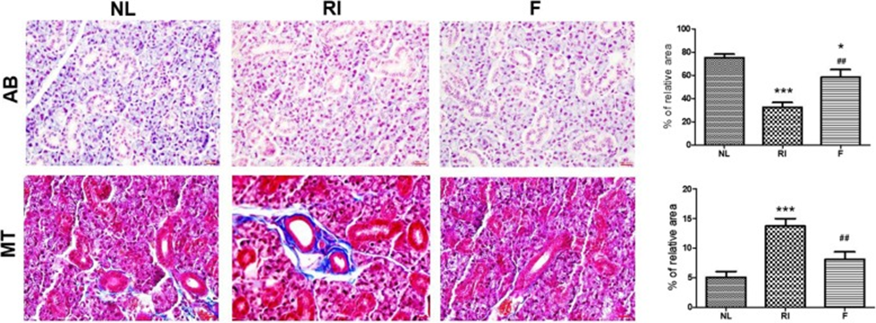
Following 12 weeks of RI administration, histological alterations within the SG were visualized using H&E, AB, and MT staining techniques. Morphometric analysis of AB staining, which indicates mucin density, showed that mucin production decreased in the RI group compared with the normal group. However, mucin production was significantly increased in the fucoidan-treated group compared with the RI group (Fig. 1, p<0.05). Through MT staining to assess the extent of fibrosis, it was observed that the RI group displayed the most significant degree of fibrosis compared to the normal group. Conversely, the fucoidan group showed a reduced level of fibrosis relative to the RI group (as can be seen in Figure 1, with a statistically significant p-value less than 0.05).
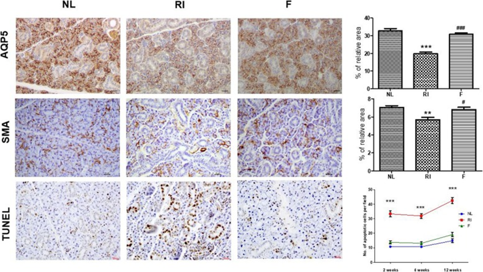
The protective action of fucoidan on salivary epithelial and myoepithelial cells was assessed using immunohistochemical staining. The expression of AQP5 (a marker for salivary epithelial cells) and α-SMA (a marker for myoepithelial cells) decreased in the RI group compared with the normal group (Figure 2, p < 0.05). The staining intensity of these cells was higher after fucoidan treatment than in the RI group, implying that fucoidan may prevent salivary epithelial and myoepithelial cells from RI-induced cytotoxicity (Figure 2, p < 0.05). TUNEL assay showed that the number of TUNEL-positive cells was significantly higher in the RI group than in the control group at 2, 4, and 12 weeks after RI, whereas it was significantly lower in the fucoidan group at 2, 4, and 12 weeks after RI (Figure 2, p < 0.05).
In the RI group, the excretion capacity of 99mTc-pertechnetate declined twelve weeks following the administration of RI, while in the fucoidan group, the excretion capacity had recuperated to a level that was equivalent to that of the normal control group.
In comparison to the RI-treated group, the fucoidan group presented with an enhanced salivary flow rate and a reduced lag time, which were observed as a result. In the fucoidan group, histological analysis of salivary gland ducts (SGs) showed mucin-rich parenchymal areas and less periductal fibrosis than in the RI-treated group. In addition, the fucoidan group showed protective effects on cells, as indicated by a greater number of salivary epithelial and myoepithelial cells than in the RI-treated group. The fucoidan group also had fewer apoptotic cells compared with the RI-treated group. The excretion of 99mTc-pertechnetate in the fucoidan group was similar to that in the control group. According to this study, administering fucoidan to mice before RI exposure reduced the SG damage that RI caused. The researchers advocate for fucoidan’s potential as a preventative measure for SG dysfunction in thyroid cancer patients receiving RI therapy.
Source: PMCID: PMC6716941 PMID: 31470847
Really interesting article! I’m always curious about what makes slots so engaging – those instant gratification mechanics are key. Thinking of trying out a new platform, maybe 777pinas download – seems geared towards quick fun for Filipino players!
Thank you for your a good offer, but I like what I am doing now.
Thank you again