
The marine-derived polysaccharide fucoidan combines multiple biological activities. Various in vitro and in vivo studies have demonstrated that fucoidan exhibits antiviral, antitumor, antioxidant, anti-inflammatory, and anticoagulant properties. In this blog, I would like to share the study, “Crude Fucoidan Extracts Impair Angiogenesis in Models Relevant for Bone Regeneration and Osteosarcoma via Reduction of VEGF and SDF-1” by Fanlu Wang et al that is considering the effects of fucoidan (fucus vesiculosus) on individual key cell types and their interactions in co-cultures via growth factors and chemokines, they assessed the effects of free fucoidan on cellular and molecular processes in angiogenesis and osteogenesis.
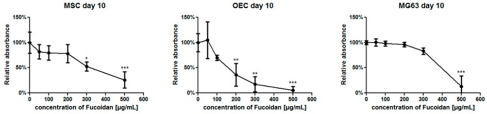
First, MTS assays were performed to investigate the potential effects of fucoidan on the metabolic activities of mesenchymal stem cell MSCs, the osteosarcoma cell line MG63, and Outgrowth endothelial cells OECs in monoculture at day 10 using different concentrations of fucoidan (Fig. 1). MTS absorbance values were presented as the relative changes between the fucoidan-treated group and the untreated control group (100%). At 100 µg/mL (Figure 1), the metabolic activity of MSCs and OECs was slightly but not significantly decreased in the fucoidan-treated group compared to the control group. At higher concentrations of fucoidan, metabolic activity was further decreased. Initial effects on OECs at a fucoidan concentration of 200 µg/mL showed that OECs appeared to be more sensitive compared to MSCs (significant effects were observed at 300 µg/mL), while MG63 appeared to be resistant to higher concentrations of fucoidan (significant effects were observed at 500 µg/mL). Based on these observations, all further experiments to evaluate angiogenesis and osteogenesis were performed at a fucoidan concentration of 100 µg/mL
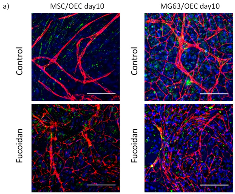
The morphology of OECs and the formation of angiogenic structures in the cocultures were observed using confocal microscopy after immunostaining with the endothelial marker VE-cadherin (Fig. 2a, shown in red). Additionally, samples were stained with the stromal-derived factor receptor CXCR4 (Fig. 2a, shown in green, with nuclear counterstain in blue). In MSC/OEC cocultures, OECs in the untreated control group exhibited elongated cell shapes and aligned into tubular structures typical of pro-angiogenic structures shown in Fig. 2a. In contrast, fewer pro-angiogenic structures were observed after fucoidan treatment (100 µg/mL), and OECs remained mainly organized as a monolayer with clear cell-cell contacts as shown by VE-cadherin staining, although the formation of angiogenic structures was not completely inhibited after fucoidan treatment. Similar to MSC/OEC cocultures, OECs in coculture with MG63 were characterized by the formation of complex vascularized structures at day 10 in the untreated control group, as shown in Fig. 2a. In the fucoidan-treated group, elongated vascularized structures of OECs were only slightly observed, mainly at the borders of OEC cell patches.
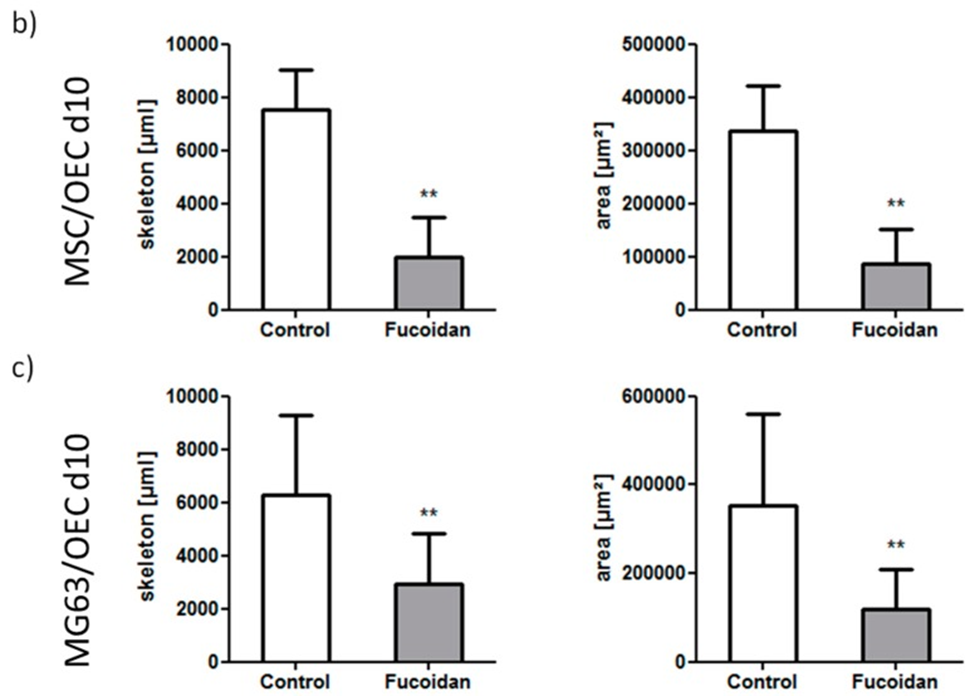
To quantify the effect of fucoidan on the formation of vascularized structures, quantitative image analysis was performed as shown in Fig. 2b, c. In both types of cocultures, the addition of fucoidan to the culture medium significantly reduced the length and area of vascularized structures, indicating the antiangiogenic properties of fucoidan in coculture models related to normal bone physiology or osteosarcoma, respectively.
To gain a better understanding of the action of fucoidan and its potential impact on cell proliferation, total DNA content was analyzed as an indicator of cell proliferation in OEC, MSC, and MG63 monocultures and both co-cultures using a Pico green-based assay. Total DNA content analyzed at day 10 was significantly decreased by fucoidan treatment in MSC monocultures but increased in OEC monocultures, whereas no significant difference was observed in MG63 monocultures. In both co-cultures, DNA content was slightly decreased in the fucoidan-treated groups, but this trend was not statistically significant in the co-cultures.
As a next step, semi-quantitative real-time PCR tests were performed on the control and fucoidan-treated monocultures and cocultures (Figure 4) to analyze the relative gene expression of molecules related to bone formation and osteoblast differentiation, factors involved in the control of angiogenesis and stem cell mobilization, and endothelial markers. In OECs, no significant effect of fucoidan treatment on the investigated genes was observed. However, a significant effect of fucoidan treatment was observed in osteoblasts. Similar effects were observed in the osteosarcoma cell line MG63, but the effect on VEGF was not significant in MG63. In addition, fucoidan treatment tentatively downregulated the pro-angiogenic factor angiopoietin 2 (Ang-2) in the cocultures. Regarding the effect of fucoidan treatment on bone differentiation markers, the results differed between MSCs and the osteosarcoma cell line MG63, showing a significant downregulation of alkaline phosphatase only in MSCs and a significant downregulation of collagen type 1 only in MG63. In the cultures of MG63 and OEC, the levels of SDF-1 and its corresponding receptor CXCR4 were decreased, again in response to fucoidan, as were collagen type 1 and angiopoietin-1.
Based on the PCR results, the amounts of angiogenesis-related factors such as VEGF, ANGPT-1, ANGPT-2, and SDF-1, which are involved in cell recruitment and osteosarcoma progression, were measured in the supernatants by ELISA to gain insight into the effect of fucoidan on protein levels. Fucoidan treatment significantly reduced VEGF levels in MSC/OEC and MG63/OEC cocultures, and similar effects were observed in MSC and MG63 monocultures. In addition, Angiopoietin-2, a pro-angiogenic factor mainly produced by endothelial cells themselves, was reduced in response to fucoidan treatment in all samples. In contrast, Angiopoietin 1, which like Angiopoietin-2 binds to the Tie-2 receptor and limits the formation of new vascular structures, but leads to stabilization of blood vessels, was increased. Finally, SDF-1, which is mainly involved in the recruitment of bone stem cells and immune cells, was significantly reduced in response to fucoidan in MG63 monocultures and both types of cocultures. These decreased protein levels of VEGF, Ang-2, and SDF-1 in co-culture in response to fucoidan are determinants of the physiological processes of angiogenesis and cell recruitment in co-culture.
To evaluate the potential effect of fucoidan on the bone formation process, mineralization levels were evaluated on day 14 of culture. MSC monocultures treated with fucoidan showed significantly decreased mineralization compared to the untreated group. Similarly, MSC/OEC cocultures showed significantly decreased mineralization in the fucoidan-treated group compared to the control group. A similar trend was observed in MG63/OEC cocultures and MG63 monocultures, but no significant effects were observed. This may be due to the tumor cell properties of MG63.
In summary, these data suggest that lower doses of fucoidan may be appropriate to limit angiogenesis, for example, in osteosarcoma.
Source: Mar Drugs. 2017 Jun 20;15(6):186. doi: 10.3390/md15060186