
Lung cancer has consistently been identified as the leading cause of cancer-related mortality in recent years. While smoking history is undeniably a key risk factor for lung cancer, it is complemented by a range of other significant contributing factors, including an individual’s lifestyle, their exposure to environmental influences, and their genetic makeup, all of which are major risk factors, and it is noteworthy that non-small cell lung cancer (NSCLC) subtypes are responsible for approximately 85% of lung cancer cases. Therefore, preventing metastasis and the spread of the tumor is extremely crucial.
Fucoidan, a polysaccharide molecule, is naturally present in brown algae. Research indicates that fucoidan possesses diverse biological functions, such as antioxidant, antibacterial, antiviral, anti-obesity, immunomodulatory, neuroprotective, and anticancer effects.
This blog post will discuss the study titled “Antimetastatic effect of fucoidan against non-small cell lung cancer by suppressing non-receptor tyrosine kinase and extracellular signal-related kinase pathway” by Nareenath Muneerungsee et al.
In order to investigate the cytotoxic impacts of fucoidan on both non-cancerous and cancerous cell lines, HaCaT cells were employed to represent normal cells, while A549 cells were utilized as a model for cancerous cells. Fucoidan at concentrations of 10–500 µg/mL was safe for normal cells, with cell viability still exceeding 90% after 48 and 72 hours of treatment. In A549 cancer cells, fucoidan at concentrations of 10–250 µg/mL significantly suppressed cell viability to less than 90% after 48 hours compared to the control. Regardless of the fucoidan concentration used, A549 cell viability stayed above 90% at both 24 and 72-hour time points post-treatment when compared to the control group.
The findings derived from the wound healing assay provided clear evidence that fucoidan acted to impede the movement of A549 lung cancer cells, as illustrated in Figure 1A. After 24 hours of treatment, the wound area gradually increased to 71.46, 69.84, 77.67, and 75.82% after 10, 25, 50, and 100 µg/mL fucoidan treatment, respectively, compared to the control group. After 48 hours of treatment with fucoidan, the percentage of the wound area significantly increased to 50.31, 56.97, 60.83, and 59.60%, respectively, compared to the control group (40.80%). The results of the wound healing assay indicated that fucoidan (10–100 µg/mL) significantly suppressed A549 lung cancer cell migration.
Phalloidin-rhodamine staining was performed to evaluate the actin cytoskeleton, including filopodia and lamellipodia, and actin stress fibers. This study’s findings indicated that fucoidan, at concentrations of 25, 50, and 100 µg/mL, decreased the fluorescence intensity. This reduction was linked to the suppression of filopodia and lamellipodia protrusion development in A549 lung cancer cells, when contrasted with cells that were not treated.
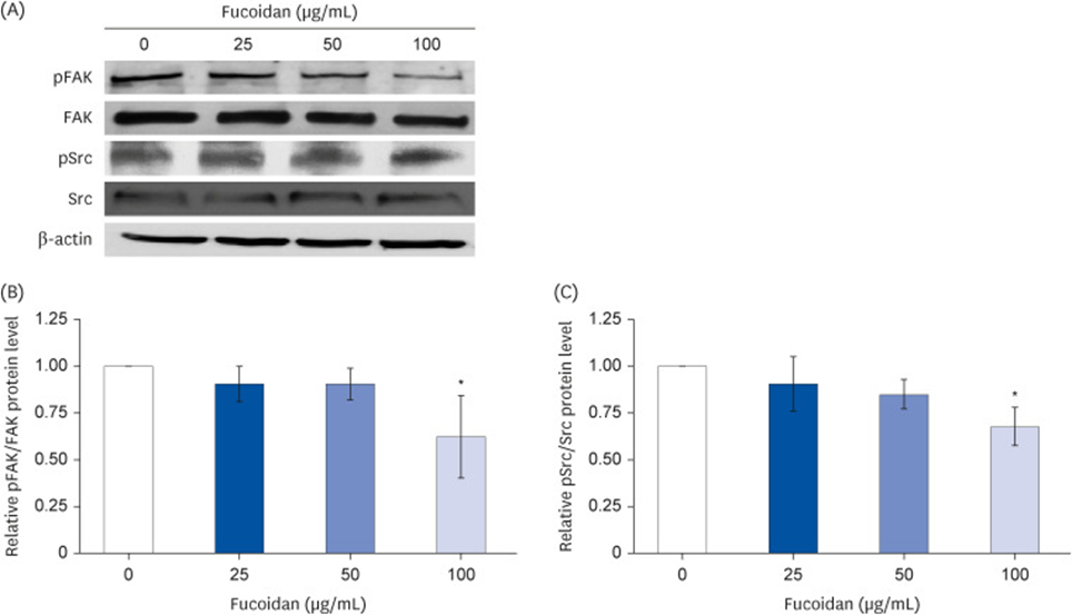
Western blot analysis was then employed to determine how fucoidan inhibits proteins involved in the process of cell migration. According to Figure 1, a concentration of 100 µg/mL of fucoidan led to a significant reduction in FAK phosphorylation (Figures 1A and 1B) and Src phosphorylation (Figures 1A and 1C) relative to the control. These results, when considered as a whole, propose that fucoidan acts to inhibit lung cancer cell migration by influencing FAK-Src signaling.
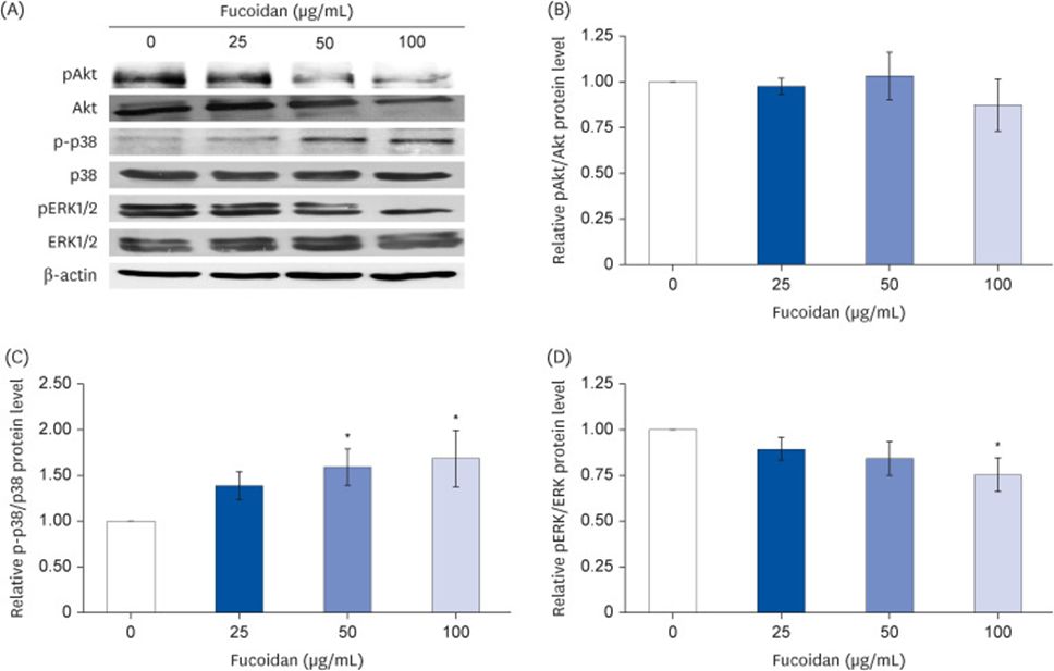
They measured the responses of the ERK1/2, p38 MAPK, and Akt signaling pathways to fucoidan treatment in A549 lung cancer cells. It was observed that fucoidan did not affect the Akt signaling pathway at any tested concentration (see Figure. 2A and B). As shown in Figure. 2A and C, Western blotting revealed that fucoidan at 50–100 µg/mL significantly increased p38 phosphorylation compared with the control group but did not alter total protein levels. Additionally, fucoidan at a concentration of 100 µg/mL demonstrably lowered ERK1/2 phosphorylation relative to the control group (see Figure. 2A and D).
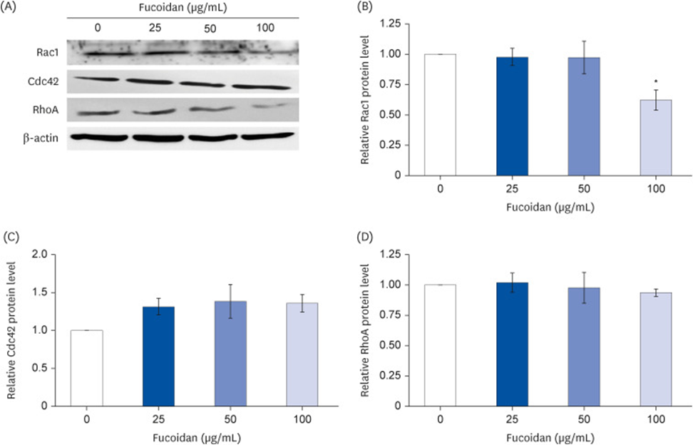
Cell migration and cytoskeletal protrusion were also regulated by downstream signaling of the Rho GTPase family. As shown in Figure 3A and B, 100 µg/mL fucoidan significantly downregulated Rac1 (relative level 0.62-fold compared to the untreated group). However, Cdc42 (Figure 3A and C) and RhoA (see Figure 3A and D) were not affected by fucoidan treatment.
The research indicates that fucoidan’s anti-cancer properties are derived from its ability to impede tumor cell mobility. The inhibition of lung cancer cell migration by fucoidan is further attributed to its suppression of the FAK, Src, and ERK1/2 signaling cascades. Additionally, the inhibition of Rac1 expression by fucoidan administration implies its participation in preventing cytoskeletal protrusion. Fucoidan appears to be a valuable compound for the future development of drugs designed to combat cancer metastasis, based on these discoveries.
Source: Nutr Res Pract. 2023 Jul 13;17(5):844–854. doi: 10.4162/nrp.2023.17.5.844