
Mitochondria are considered the cellular energy source. In dopaminergic neurons of Parkinson’s disease (PD), mitochondria are the organelles most susceptible to damage. Despite the importance of protecting neuromitochondrial function in PD patients, the detailed mechanisms of mitochondrial dysfunction during the pathogenesis and pathophysiological progression of PD remain unclear.
In this blog, I would like to inform you of the study, “Fucoidan Suppresses Mitochondrial Dysfunction and Cell Death against 1-Methyl-4-Phenylpyridinum-Induced Neuronal Cytotoxicity via Regulation of PGC-1α Expression” by Yong-Seok Han et al which investigated the protective effect of fucoidan on mitochondria in SH-SY5Y neuronal cells induced by 1-methyl-4-phenylpyridinium (MPP+), a neurotoxin used as a PD model.
Fucoidan (Fucus vesiculosus) is a sulfated polysaccharide extracted from brown algae. Numerous studies have reported its biological activities, including antitumor, anti-inflammatory, and antioxidant properties. Furthermore, fucoidan enhances stem cell physiological activity and therapeutic efficacy.
To investigate the protective effect of fucoidan against 1-methyl-4-phenyl-pyridinium (MPP+)- induced oxidative stress-induced cytotoxicity, neuroblastoma cell line (SH-SY5Y cells) were first treated with various concentrations of fucoidan. Fucoidan at 50 μg/mL stimulated cell proliferation after 24 hours. Furthermore, SH-SY5Y cells were treated with MPP+ at predetermined doses (0, 0.1, 0.5, 1, and 2 mM). The MPP+-induced decrease in SH-SY5Y cell viability was greatest at 2 mM. However, the decrease in viability due to MPP+-induced apoptosis was suppressed by fucoidan pretreatment. To further investigate the protective effect of fucoidan against MPP+-induced senescence in SH-SY5Y cells, they performed Western blotting to quantify SMP30 levels and a senescence-associated β-galactosidase assay. However, these results indicated that MPP+ did not induce senescence in SH-SY5Y cells. These results suggest that fucoidan protects SH-SY5Y cells from MPP+-induced apoptosis. These findings suggest that fucoidan improves the proliferation and cell viability of SH-SY5Y cells in the MPP+-induced PD model.
To investigate the effect of fucoidan on oxidative stress in response to MPP+ treatment, MPP+-treated cells were incubated with dihydroethidium (DHE) to detect reactive oxygen species (ROS) and measured changes in oxidative stress levels using fluorescence microscopy imaging. SH-SY5Y cells exposed to 2 mM MPP+ for 24 h showed significantly elevated oxidative stress levels compared with untreated cells, indicating that MPP+ enhances oxidative stress (Fig. 1A, B). Fluorescence microscopy imaging of DHE revealed that fucoidan treatment significantly suppressed the increase in oxidative stress levels (Fig. 1A, B). Furthermore, to investigate whether MPP+ reduces mitochondrial membrane potential through oxidative stress, they measured the activity of complexes I and IV (Fig. 1C, D) and the mitochondrial oxygen consumption rate (Fig. 1E). The results show that MPP+ reduced the activity of complexes I and IV and the mitochondrial oxygen consumption rate. In contrast, fucoidan pretreatment protected the activity of complexes I and IV and the mitochondrial oxygen consumption rate from MPP+ (Fig. 1C–E). However, in the absence of MPP+, fucoidan did not alter the oxygen consumption rate of SH-SY5Y cells, suggesting that fucoidan may affect mitochondrial oxidative phosphorylation under MPP+-induced stress conditions. These results suggest that fucoidan protects SH-SY5Y cells from MPP+-induced oxidative stress and mitochondrial dysfunction.
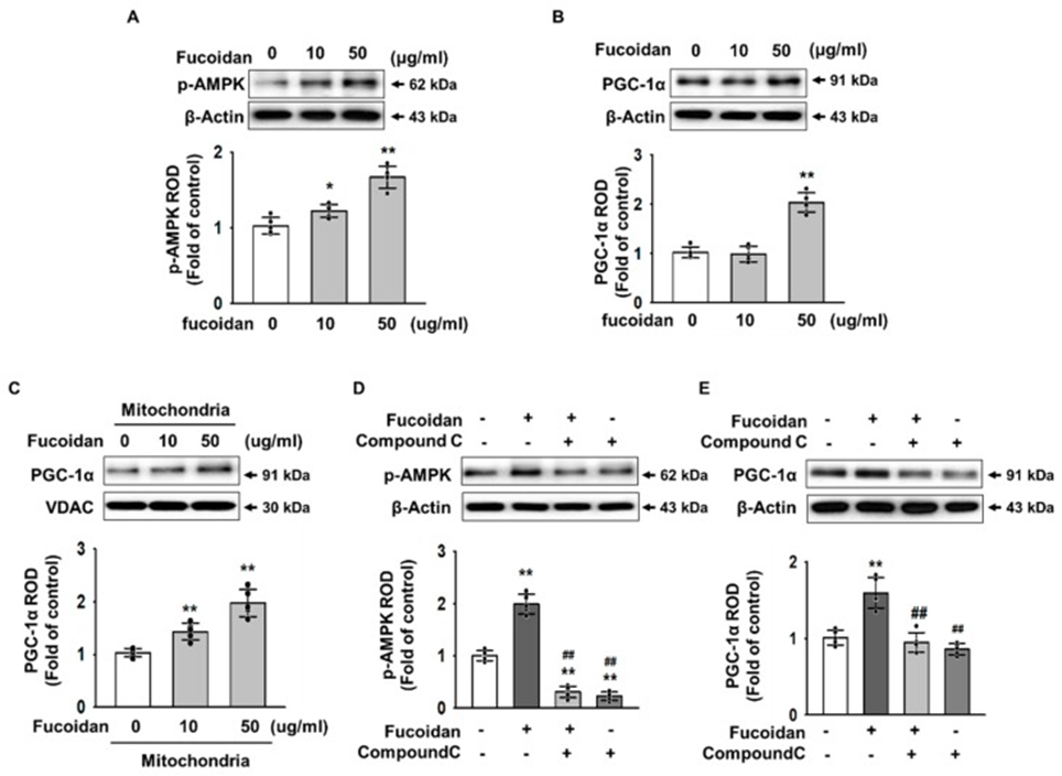
Next, to investigate the key mediators of mitochondrial function protected by fucoidan, they analyzed the expression of 5′ adenosine monophosphate-activated protein kinase (AMPK) and PGC-1α in a concentration-dependent manner (0-50 μg/mL) by Western blot analysis. Western blot analysis showed that AMPK phosphorylation was increased in SH-SY5Y cells treated with fucoidan (50 μg/mL) (Figure 2A). PGC-1α expression was also increased at the same concentration (Figure 2B). Furthermore, PGC-1α entry into mitochondria was increased (Figure 2C). Furthermore, using the AMPK inhibitor compound C, they confirmed that fucoidan activates PGC-1α by regulating AMPK phosphorylation. When AMPK phosphorylation was inhibited, fucoidan did not increase PGC-1α expression in SH-SY5Y cells (Figure 2D, E). These results suggest that fucoidan regulates the expression of PGC-1α, a key factor in mitochondrial biogenesis, through the phosphorylation of AMPK.
To clarify whether the reduced mitochondrial membrane potential was caused by a decrease in expression of PGC-1α, we measured complex I & IV activities and mitochondrial O2 consumption ratio. As a result, when the expression of PGC-1α was inhibited, the fucoidan did not increase the activities of complex I & IV (Figure 3A, B), and the O2 consumption ratio of the mitochondria was also inhibited by inhibiting the expression of PGC-1α (Figure 3C). These results indicated that the effect of fucoidan reduced dysfunctional mitochondria by increasing PGC-1α expression.
To investigate the effect of fucoidan on MPP+-induced apoptosis, we analyzed the expression levels of B-cell lymphoma 2 (BCL2), BCL-2-like protein 4 (Bax), cleaved poly (ADP-ribose) polymerase 1 (PARP-1), and cleaved caspase 3, which are associated with cell survival or apoptosis, by Western blot assay. When MPP+-induced oxidative stress induced apoptosis in SH-SY5Y cells, fucoidan increased the expression of BCL2, but significantly decreased the expression levels of Bax, cleaved PARP-1, and cleaved caspase 3. Furthermore, apoptosis was assessed using propidium iodide (PI)/annexin V staining. Fucoidan reduced apoptosis via regulating PGC-1α expression/ These results suggest that fucoidan inhibits apoptosis via increasing PGC-1α expression.
In conclusion, the study revealed that regulating the AMPK-PGC-1α pathway, fucoidan from Fucus vesiculosus, protects against mitochondrial dysfunction and apoptosis in a PD cell model exhibiting MPP+-induced mitochondrial dysfunction, high oxidative stress, and decreased survival rate. These findings suggest that fucoidan may be used to enhance the efficacy of therapeutic strategies against neurotoxicity insults in PD.


Source: Mar Drugs. 2019 Sep 2;17(9):518. doi: 10.3390/md17090518